WB
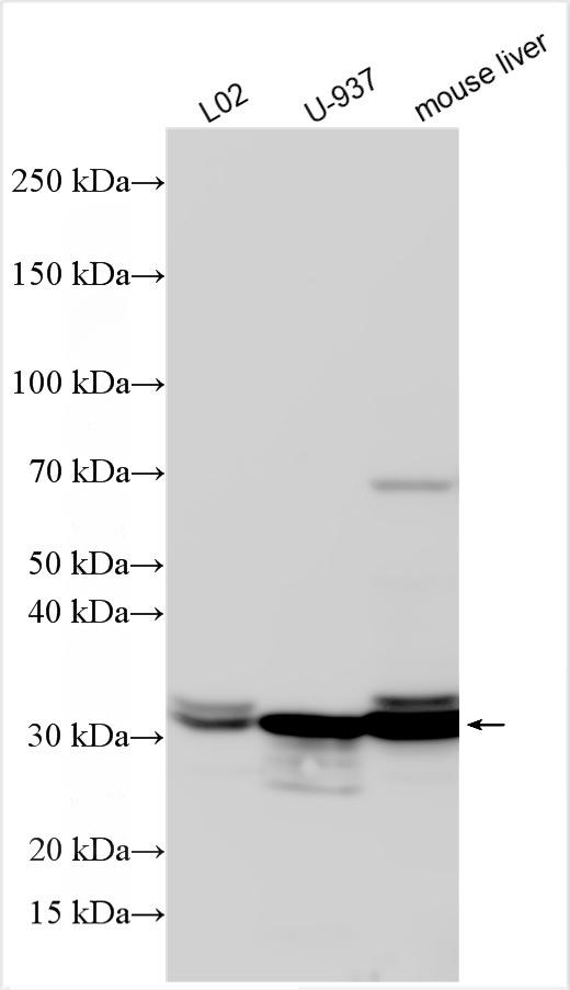
Various lysates were subjected to SDS PAGE followed by western blot with SULT1A1 antibody at dilution of 1:3000 incubated at room temperature for 1.5 hours.WB
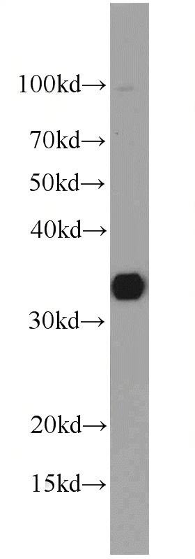
mouse liver tissue were subjected to SDS PAGE followed by western blot with SULT1A1 antibody at dilution of 1:1000 incubated at room temperature for 1.5 hours.IHC
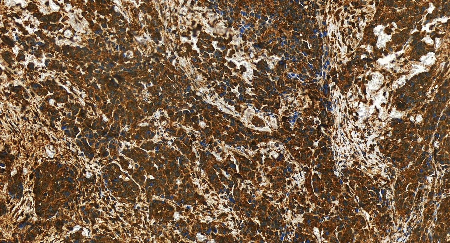
Immunohistochemical analysis of paraffin-embedded human ovary cancer tissue slide using SULT1A1 antibody at dilution of 1:400 (under 20x lens). Heat mediated antigen retrieval with Tris-EDTA buffer (pH 9.0).IP
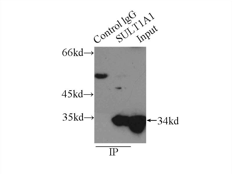
IP result of anti-SULT1A1 (IP: 3ug; Detection: 1:1000) with mouse liver tissue lysate 12500ug.ICC/IF
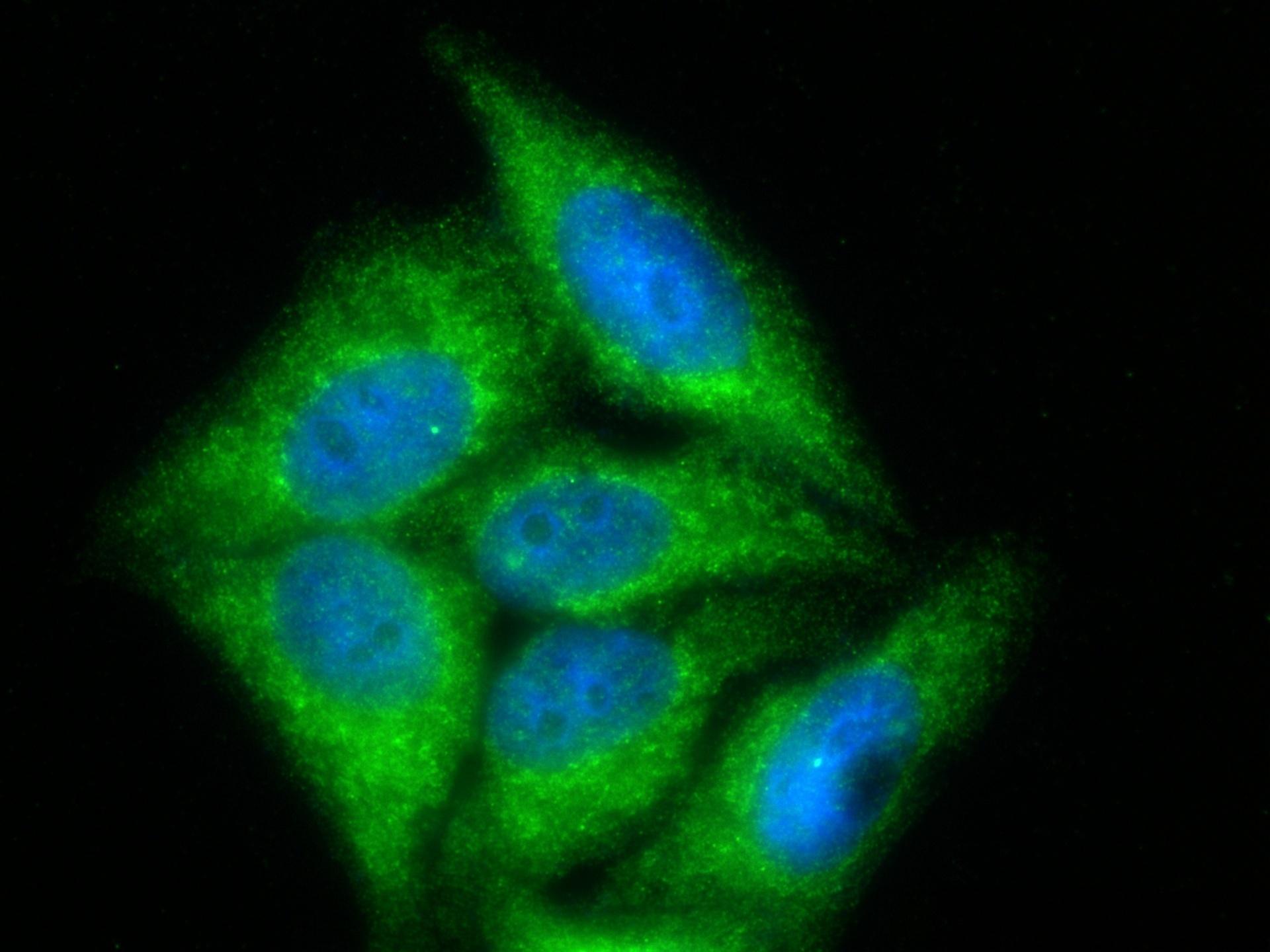
Immunofluorescent analysis of (-20°C Ethanol) fixed HepG2 cells using SULT1A1 antibody at dilution of 1:400 and CoraLite®488-Conjugated Goat Anti-Rabbit IgG(H+L) .FC
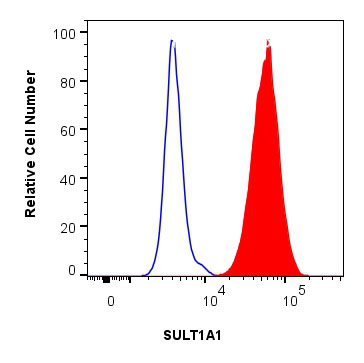
1x10^6 HepG2 cells were intracellularly stained with 0.25 ug SULT1A1 Polyclonal antibody and CoraLite®488-Conjugated Goat Anti-Rabbit IgG(H+L) (red), or 0.25 ug rabbit IgG isotype control (blue). Cells were fixed with 4% PFA and permeabilized with Flow Cytometry Perm Buffer.| Product Name | SULT1A1 Polyclonal antibody |
|---|---|
| Antibody Type | Primary Antibodies |
| Immunogen | Recombinant human SULT1A1 |
| Clonality | Polyclonal |
|---|---|
| Isotype | IgG |
| Host Species | Rabbit |
| Tested Applications | FCICC/IFIHCIPWB |
| WB:1:1000-1:6000 ICC/IF:1:200-1:800 IHC:1:200-1:800 IP:0.5-4.0 ug for 1.0-3.0 mg of total protein lysate FC: 0.25 ug per 10^6 cells in a 100 µl suspension |
|
| Species Reactivity | HumanMouseRat |
| Concentration | 1mg/ml |
| Purification | Affinity purified |
| Gene Symbol | SULT1A1 |
|---|---|
| Gene Synonyms | PST STP STP1 P-PST ST1A1 ST1A3 TSPST1 ts-PST P-PST 1 HAST1/HAST2 |
| Gene Full Name | sulfotransferase family 1A member 1 |
| Gene Summary | Sulfotransferase enzymes catalyze the sulfate conjugation of many hormones, neurotransmitters, drugs, and xenobiotic compounds. These cytosolic enzymes are different in their tissue distributions and substrate specificities. The gene structure (number and length of exons) is similar among family members. This gene encodes one of two phenol sulfotransferases with thermostable enzyme activity. Multiple alternatively spliced variants that encode two isoforms have been identified for this gene. [provided by RefSeq, Jul 2008] |
| Alternative Names | EC:2.8.2.1 HAST1/HAST2 OK/SW-cl.88 P PST P PST 1 |
| Molecular Weight(MW) | 34 kDa |
| Cellular Localization | Cytoplasm |
WB
Various lysates were subjected to SDS PAGE followed by western blot with SULT1A1 antibody at dilution of 1:3000 incubated at room temperature for 1.5 hours.
WB
mouse liver tissue were subjected to SDS PAGE followed by western blot with SULT1A1 antibody at dilution of 1:1000 incubated at room temperature for 1.5 hours.
IHC
Immunohistochemical analysis of paraffin-embedded human ovary cancer tissue slide using SULT1A1 antibody at dilution of 1:400 (under 20x lens). Heat mediated antigen retrieval with Tris-EDTA buffer (pH 9.0).
IP
IP result of anti-SULT1A1 (IP: 3ug; Detection: 1:1000) with mouse liver tissue lysate 12500ug.
ICC/IF
Immunofluorescent analysis of (-20°C Ethanol) fixed HepG2 cells using SULT1A1 antibody at dilution of 1:400 and CoraLite®488-Conjugated Goat Anti-Rabbit IgG(H+L) .
FC
1x10^6 HepG2 cells were intracellularly stained with 0.25 ug SULT1A1 Polyclonal antibody and CoraLite®488-Conjugated Goat Anti-Rabbit IgG(H+L) (red), or 0.25 ug rabbit IgG isotype control (blue). Cells were fixed with 4% PFA and permeabilized with Flow Cytometry Perm Buffer.| Application Notes | WB:1:1000-1:6000 ICC/IF:1:200-1:800 IHC:1:200-1:800 IP:0.5-4.0 ug for 1.0-3.0 mg of total protein lysate FC: 0.25 ug per 10^6 cells in a 100 µl suspension |
|---|
| Form | Liquid |
|---|---|
| Storage Instructions | Store at -20°C. Stable for one year after shipment. |
| Storage Buffer | PBS with 0.02% sodium azide and 50% glycerol pH 7.3. |


