WB
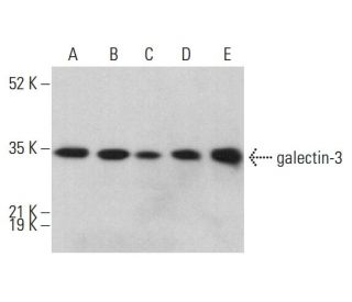
Western blot analysis of galectin-3 expression in nuclear extracts of HeLa (A) and SW480 (B), as well as whole cell lysates of A-431 (C), MCF7 (D) and HeLa (E). Detection reagent: m-IgGκ BP-HRP.IHC
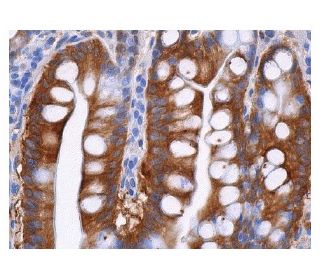
Immunoperoxidase staining of formalin-fixed, paraffin-embedded human small intestine tissue, showing cytoplasmic and membrane staining in glandular cells.WB
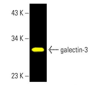
Direct fluorescent Western blot analysis of galectin-3 expression in 3611-RF whole cell lysates. Blocking was performed with UltraCruz® Blocking ReagentIHC
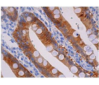
Immunoperoxidase staining of formalin-fixed, paraffin-embedded human duodenum tissue exhibits cytoplasmic staining in glandular cells.| Product Name | Mouse Monoclonal antibody to galectin-3 |
|---|---|
| Antibody Type | Primary Antibodies |
| Product description | Firefly Luciferase Antibody is a mouse monoclonal IgG2a κ antibody against luciferase (also referred to as anti-firefly luciferase antibody). It enables the detection of luciferase protein derived from Photinus pyralis via Western Blot (WB), Immunoprecipitation (IP), Immunofluorescence (IF) and Enzyme-Linked Immunosorbent Assay (ELISA).
This anti-luciferase antibody is available in both unconjugated form and a variety of conjugated formats, including agarose, horseradish peroxidase (HRP), phycoerythrin (PE), fluorescein isothiocyanate (FITC), and multiple Alexa Fluor® conjugates.
Luciferase isolated from Photinus pyralis, a common North American firefly, is one of the most extensively studied enzymes that catalyze light production in bioluminescent organisms. As a member of the ATP-dependent AMP-binding enzyme family, it emits green light at a wavelength of 562 nm.
The chemical reaction catalyzed by luciferase is as follows: Photinus luciferin + O₂ + ATP → Oxidized Photinus luciferin + CO₂ + AMP + Pyrophosphate + Light |
| Immunogen | raised against full length galectin-3 of human origin. |
| Clonality | Monoclonal |
|---|---|
| Isotype | IgG1, k |
| Host Species | Mouse |
| Tested Applications | IHCWB |
| WB:1:100-1:1000 IHC:1:50-1:500 |
|
| Species Reactivity | HumanMouseRat |
| Concentration | 0.1mg/ml |
| Purification | Affinity purified |
| Gene Symbol | LGALS3 |
|---|---|
| Gene Synonyms | L31 GAL3 MAC2 CBP35 GALBP GALIG LGALS2 |
| Gene Full Name | galectin 3 |
| Gene Summary | This gene encodes a member of the galectin family of carbohydrate binding proteins. Members of this protein family have an affinity for beta-galactosides. The encoded protein is characterized by an N-terminal proline-rich tandem repeat domain and a single C-terminal carbohydrate recognition domain. This protein can self-associate through the N-terminal domain allowing it to bind to multivalent saccharide ligands. This protein localizes to the extracellular matrix, the cytoplasm and the nucleus. This protein plays a role in numerous cellular functions including apoptosis, innate immunity, cell adhesion and T-cell regulation. The protein exhibits antimicrobial activity against bacteria and fungi. Alternate splicing results in multiple transcript variants.[provided by RefSeq, Oct 2014] |
| Molecular Weight(MW) | 31 kDa |
| Cellular Localization | Cytoplasm,Nucleus,Secreted,Spliceosome |
WB
Western blot analysis of galectin-3 expression in nuclear extracts of HeLa (A) and SW480 (B), as well as whole cell lysates of A-431 (C), MCF7 (D) and HeLa (E). Detection reagent: m-IgGκ BP-HRP.
IHC
Immunoperoxidase staining of formalin-fixed, paraffin-embedded human small intestine tissue, showing cytoplasmic and membrane staining in glandular cells.
WB
Direct fluorescent Western blot analysis of galectin-3 expression in 3611-RF whole cell lysates. Blocking was performed with UltraCruz® Blocking Reagent
IHC
Immunoperoxidase staining of formalin-fixed, paraffin-embedded human duodenum tissue exhibits cytoplasmic staining in glandular cells.| Application Notes | WB:1:100-1:1000 IHC:1:50-1:500 |
|---|
| Form | Liquid |
|---|---|
| Storage Instructions | Store at 4° C, **DO NOT FREEZE**. Stable for one year from the date of shipment. Non-hazardous. No MSDS required. |
| Storage Buffer | Each vial contains 200 µg IgG1 kappa light chain in 1.0 ml of PBS with < 0.1% sodium azide and 0.1% gelatin. |


