WB
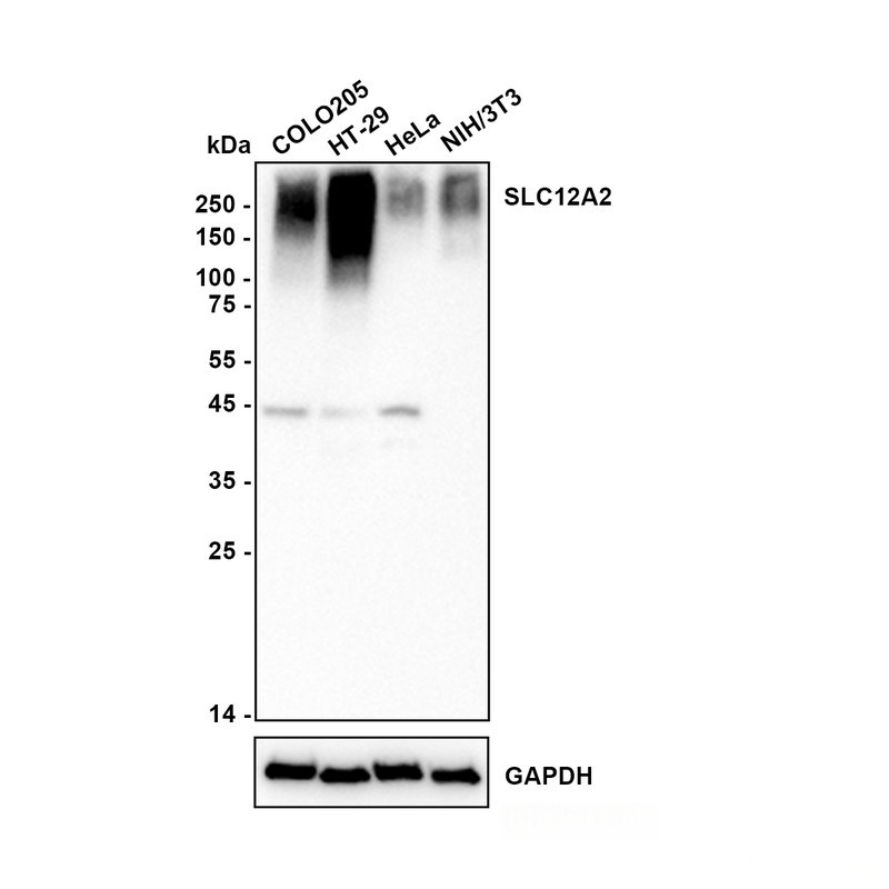
Western blot analysis of SLC12A2 / NKCC1 on different lysates with Rabbit anti-SLC12A2 / NKCC1 antibody at 1/5,000 dilution. Lysates/proteins at 20 µg/Lane. Exposure time: 20 seconds; Blocking: 5% NFDM/TBST, 1 hour at room temperature Primary antibody: Rabbit anti-SLC12A2 / NKCC1 antibody,1/5,000 in primary antibody dilution buffer, overnight at 4 °C Secondary antibody: Goat anti-Rabbit IgG-HRP , 1/50,000 in 5% NFDM/TBST, 1 hour at room temperature.WB
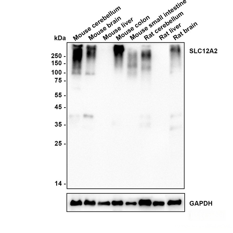
Western blot analysis of SLC12A2 / NKCC1 on different lysates with Rabbit anti-SLC12A2 / NKCC1 antibody at 1/5,000 dilution. Lysates/proteins at 20 µg/Lane. Exposure time: 20 seconds; Low expression: Liver (PMID:12000719) Blocking: 5% NFDM/TBST, 1 hour at room temperature Primary antibody: Rabbit anti-SLC12A2 / NKCC1, 1/5,000 in primary antibody dilution buffer, overnight at 4 °C Secondary antibody: Goat anti-Rabbit IgG-HRP, 1/50,000 in 5% NFDM/TBST, 1 hour at room temperature.ICC/IF
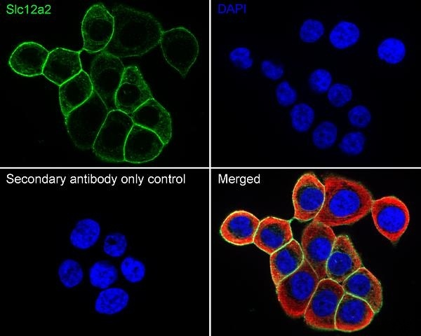
Species: Human Sample: HT-29 (Human colorectal adenocarcinoma cells) Fixation: 4% Paraformaldehyde, 15 minutes at room temperature. Permeabilization: 0.1% Triton X-100, 15 minutes at room temperature. Blocking: 1% BSA + 10% normal goat serum, 1 hour at room temperature. Antibody dilution buffer: 1% BSA in PBST. Primary antibody: Rabbit anti-SLC12A2 / NKCC1 antibody, 1/500, overnight at 4°C. Secondary antibody: Goat Anti-Rabbit IgG (488), 45 minutes at room temperature. Counterstain: Beta tubulin (red), 1/100, overnight at 4℃. The Nuclear counterstain was DAPI (Blue).IF-F
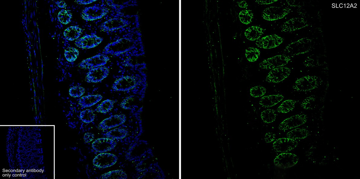
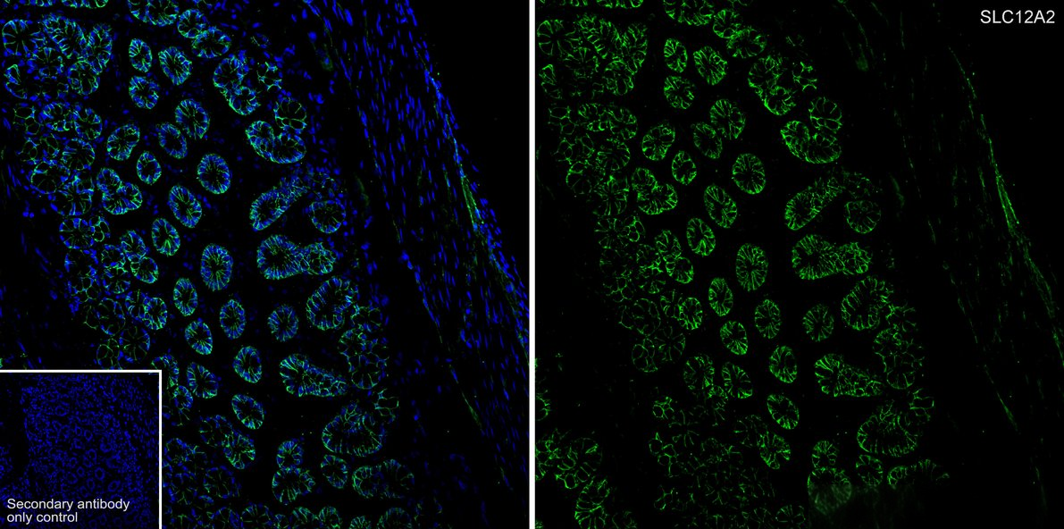
Species: Mouse, Tissue: Colon, Sample: Frozen section, Antigen retrieval: Not required, Wash buffer: 1× TBST, Blocking: 10% normal goat serum + 0.5 % Triton X-100 + 0.3 M Glycine in PBS, 10 minutes at room temperature. Primary antibody:Rabbit anti-SLC12A2 / NKCC1 antibody, 1/500, overnight at 4°C. Secondary antibody: Goat Anti-Rabbit IgG (488), 1.5 hours at room temperature.IF-F
Species: Rat Tissue: Colon Sample: Frozen section Antigen retrieval: Not required Wash buffer: 1× TBST Blocking: 10% normal goat serum + 0.5 % Triton X-100 + 0.3 M Glycine in PBS, 10 minutes at room temperature. Primary antibody: Rabbit anti-SLC12A2 / NKCC1 antibody, 1/500, overnight at 4°C. Secondary antibody: Goat Anti-Rabbit IgG (488), 1.5 hours at room temperature.IHC
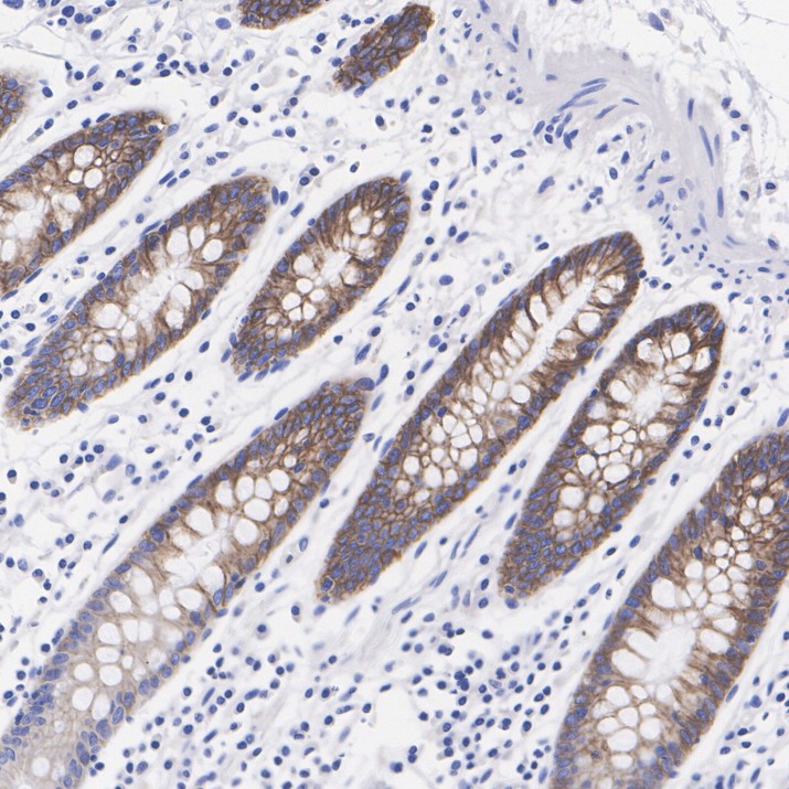
Species: Human Tissue: Colon Sample: Paraffin-embedded section Antigen retrieval: Heat-mediated, Tris-EDTA buffer (pH 9.0), 20 minutes at 95°C. Wash buffer: 1× TBST Endogenous peroxidase blocking: 3% H₂O₂, 10 minutes at room temperature. Blocking: 1% BSA + 10% normal goat serum, 10 minutes at room temperature. Primary antibody: 1/5,000, 1 hour at room temperature. Secondary antibody: 20 minutes at room temperature.IHC
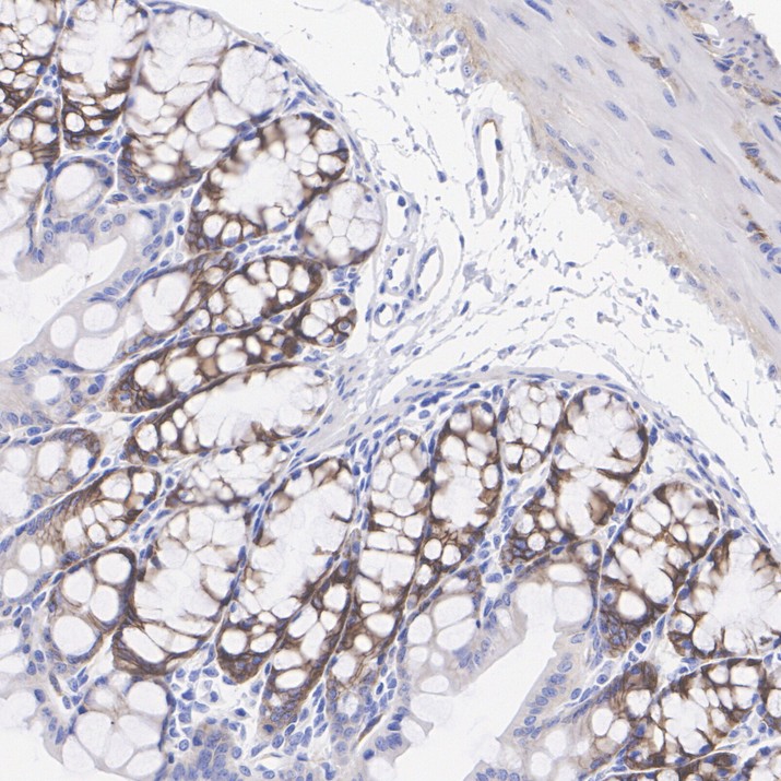
Species: Mouse Tissue: Colon Sample: Paraffin-embedded section Antigen retrieval: Heat-mediated, Tris-EDTA buffer (pH 9.0), 20 minutes at 95°C. Wash buffer: 1× TBST Endogenous peroxidase blocking: 3% H₂O₂, 10 minutes at room temperature. Blocking: 1% BSA + 10% normal goat serum, 10 minutes at room temperature. Primary antibody: 1/5,000, 1 hour at room temperature. Secondary antibody: 20 minutes at room temperature.IHC
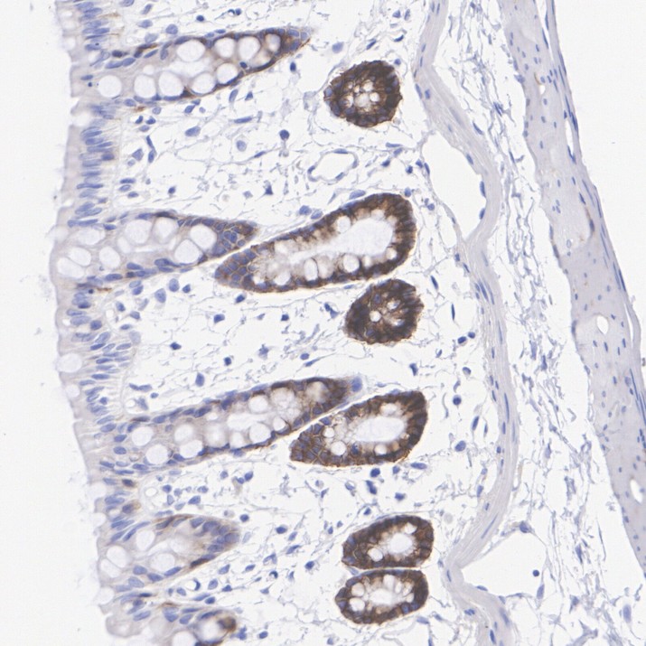
Species: Rat Tissue: Colon Sample: Paraffin-embedded section Antigen retrieval: Heat-mediated, Tris-EDTA buffer (pH 9.0), 20 minutes at 95°C. Wash buffer: 1× TBST Endogenous peroxidase blocking: 3% H₂O₂, 10 minutes at room temperature. Blocking: 1% BSA + 10% normal goat serum, 10 minutes at room temperature. Primary antibody: 1/5,000, 1 hour at room temperature. Secondary antibody: 20 minutes at room temperature.FC
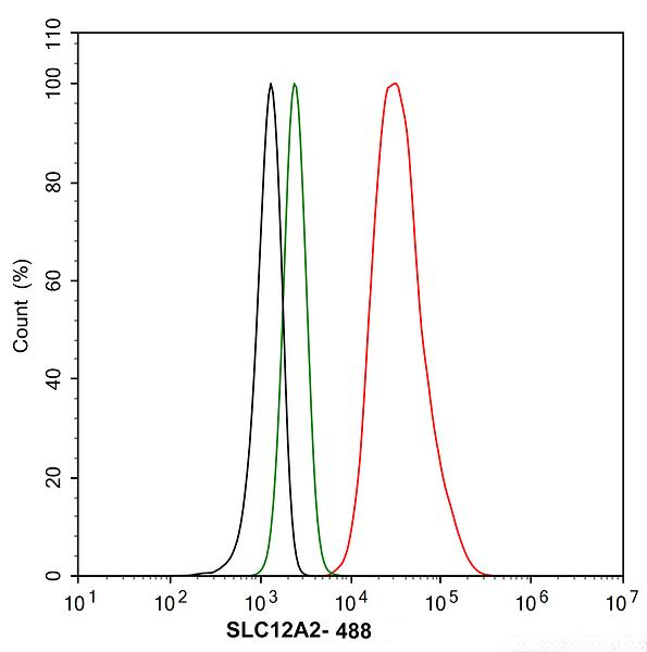
Species: Human Sample: HT-29 (Human colorectal adenocarcinoma cells) Fixation: 4% Paraformaldehyde, 15 minutes at room temperature. Permeabilization: 1x Permeabilization Buffer (eBioscience), 15 minutes at room temperature. Blocking: 1% BSA + 10% normal goat serum, 15 minutes at room temperature. Antibody dilution buffer: 1x PBS. Primary antibody: Rabbit anti-SLC12A2 / NKCC1 antibody (1/1,000) (Red) compared with Rabbit IgG Isotype Control (Green), 15 minutes at room temperature. Secondary antibody: Goat Anti-Rabbit IgG (488), 15 minutes at room temperature.IP
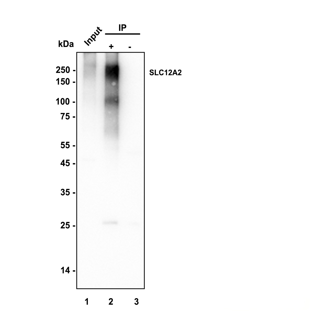
SLC12A2 / NKCC1 was immunoprecipitated in 0.2 mg COLO205 (Human colon adenocarcinoma cells) cell lysate with Rabbit anti-SLC12A2 / NKCC1 antibody at 2 µg/10 µl beads. Western blot was performed from the immunoprecipitate using Rabbit anti-SLC12A2 / NKCC1 antibody at 1/5,000 dilution. Anti-Rabbit IgG for IP Nano-secondary antibody (NBI01H) at 1/5,000 dilution was used for 1 hour at room temperature. Lane 1: COLO205 cell lysate (input) Lane 2: IP in COLO205 cell lysate Lane 3: Rabbit IgG instead of SLC12A2 / NKCC1 antibody in COLO205 cell lysate Exposure time: 7 seconds Blocking: 5% NFDM/TBST, 1 hour at room temperature Primary dilution: 1/5,000 in primary antibody dilution buffer, 2 hours at room temperature.| Product Name | SLC12A2 / NKCC1 Recombinant Rabbit Monoclonal Antibody |
|---|---|
| Antibody Type | Primary Antibodies |
| Immunogen | Recombinant protein within mouse SLC12A2 / NKCC1 aa 101-279. |
| Clonality | Monoclonal |
|---|---|
| Isotype | IgG |
| Host Species | Rabbit |
| Tested Applications | FCICC/IFIF-FIHCIPWB |
| WB:1:5000 ICC/IF:1:500 IF-F:1:500 IHC:1:5000 FC:1:1000 IP:1-2μg/sample |
|
| Species Reactivity | HumanMouseRat |
| Concentration | 1mg/ml |
| Purification | Protein A |
| Gene Symbol | SLC12A2 |
|---|---|
| Gene Synonyms | BSC BSC2 CCC1 BSC-2 KILQS NKCC1 hNKCC1 PPP1R141 |
| Gene Full Name | solute carrier family 12 member 2 |
| Gene Summary | The protein encoded by this gene mediates sodium and chloride transport and reabsorption. The encoded protein is a membrane protein and is important in maintaining proper ionic balance and cell volume. This protein is phosphorylated in response to DNA damage. Three transcript variants encoding two different isoforms have been found for this gene. [provided by RefSeq, Jan 2012] |
| Alternative Names | Solute carrier family 12 member 2 Basolateral Na-K-Cl symporter Bumetanide-sensitive sodium-(potassium)-chloride cotransporter 2 (BSC2) Na-K-2Cl cotransporter 1 (hNKCC1) SLC12A2 NKCC1 |
| Molecular Weight(MW) | 131 kDa(Observed band size: 150-200 kDa) |
| Cellular Localization | Basolateral cell membrane. |
WB
Western blot analysis of SLC12A2 / NKCC1 on different lysates with Rabbit anti-SLC12A2 / NKCC1 antibody at 1/5,000 dilution. Lysates/proteins at 20 µg/Lane. Exposure time: 20 seconds; Blocking: 5% NFDM/TBST, 1 hour at room temperature Primary antibody: Rabbit anti-SLC12A2 / NKCC1 antibody,1/5,000 in primary antibody dilution buffer, overnight at 4 °C Secondary antibody: Goat anti-Rabbit IgG-HRP , 1/50,000 in 5% NFDM/TBST, 1 hour at room temperature.
WB
Western blot analysis of SLC12A2 / NKCC1 on different lysates with Rabbit anti-SLC12A2 / NKCC1 antibody at 1/5,000 dilution. Lysates/proteins at 20 µg/Lane. Exposure time: 20 seconds; Low expression: Liver (PMID:12000719) Blocking: 5% NFDM/TBST, 1 hour at room temperature Primary antibody: Rabbit anti-SLC12A2 / NKCC1, 1/5,000 in primary antibody dilution buffer, overnight at 4 °C Secondary antibody: Goat anti-Rabbit IgG-HRP, 1/50,000 in 5% NFDM/TBST, 1 hour at room temperature.
ICC/IF
Species: Human Sample: HT-29 (Human colorectal adenocarcinoma cells) Fixation: 4% Paraformaldehyde, 15 minutes at room temperature. Permeabilization: 0.1% Triton X-100, 15 minutes at room temperature. Blocking: 1% BSA + 10% normal goat serum, 1 hour at room temperature. Antibody dilution buffer: 1% BSA in PBST. Primary antibody: Rabbit anti-SLC12A2 / NKCC1 antibody, 1/500, overnight at 4°C. Secondary antibody: Goat Anti-Rabbit IgG (488), 45 minutes at room temperature. Counterstain: Beta tubulin (red), 1/100, overnight at 4℃. The Nuclear counterstain was DAPI (Blue).
IF-F
Species: Mouse, Tissue: Colon, Sample: Frozen section, Antigen retrieval: Not required, Wash buffer: 1× TBST, Blocking: 10% normal goat serum + 0.5 % Triton X-100 + 0.3 M Glycine in PBS, 10 minutes at room temperature. Primary antibody:Rabbit anti-SLC12A2 / NKCC1 antibody, 1/500, overnight at 4°C. Secondary antibody: Goat Anti-Rabbit IgG (488), 1.5 hours at room temperature.
IF-F
Species: Rat Tissue: Colon Sample: Frozen section Antigen retrieval: Not required Wash buffer: 1× TBST Blocking: 10% normal goat serum + 0.5 % Triton X-100 + 0.3 M Glycine in PBS, 10 minutes at room temperature. Primary antibody: Rabbit anti-SLC12A2 / NKCC1 antibody, 1/500, overnight at 4°C. Secondary antibody: Goat Anti-Rabbit IgG (488), 1.5 hours at room temperature.
IHC
Species: Human Tissue: Colon Sample: Paraffin-embedded section Antigen retrieval: Heat-mediated, Tris-EDTA buffer (pH 9.0), 20 minutes at 95°C. Wash buffer: 1× TBST Endogenous peroxidase blocking: 3% H₂O₂, 10 minutes at room temperature. Blocking: 1% BSA + 10% normal goat serum, 10 minutes at room temperature. Primary antibody: 1/5,000, 1 hour at room temperature. Secondary antibody: 20 minutes at room temperature.
IHC
Species: Mouse Tissue: Colon Sample: Paraffin-embedded section Antigen retrieval: Heat-mediated, Tris-EDTA buffer (pH 9.0), 20 minutes at 95°C. Wash buffer: 1× TBST Endogenous peroxidase blocking: 3% H₂O₂, 10 minutes at room temperature. Blocking: 1% BSA + 10% normal goat serum, 10 minutes at room temperature. Primary antibody: 1/5,000, 1 hour at room temperature. Secondary antibody: 20 minutes at room temperature.
IHC
Species: Rat Tissue: Colon Sample: Paraffin-embedded section Antigen retrieval: Heat-mediated, Tris-EDTA buffer (pH 9.0), 20 minutes at 95°C. Wash buffer: 1× TBST Endogenous peroxidase blocking: 3% H₂O₂, 10 minutes at room temperature. Blocking: 1% BSA + 10% normal goat serum, 10 minutes at room temperature. Primary antibody: 1/5,000, 1 hour at room temperature. Secondary antibody: 20 minutes at room temperature.
FC
Species: Human Sample: HT-29 (Human colorectal adenocarcinoma cells) Fixation: 4% Paraformaldehyde, 15 minutes at room temperature. Permeabilization: 1x Permeabilization Buffer (eBioscience), 15 minutes at room temperature. Blocking: 1% BSA + 10% normal goat serum, 15 minutes at room temperature. Antibody dilution buffer: 1x PBS. Primary antibody: Rabbit anti-SLC12A2 / NKCC1 antibody (1/1,000) (Red) compared with Rabbit IgG Isotype Control (Green), 15 minutes at room temperature. Secondary antibody: Goat Anti-Rabbit IgG (488), 15 minutes at room temperature.
IP
SLC12A2 / NKCC1 was immunoprecipitated in 0.2 mg COLO205 (Human colon adenocarcinoma cells) cell lysate with Rabbit anti-SLC12A2 / NKCC1 antibody at 2 µg/10 µl beads. Western blot was performed from the immunoprecipitate using Rabbit anti-SLC12A2 / NKCC1 antibody at 1/5,000 dilution. Anti-Rabbit IgG for IP Nano-secondary antibody (NBI01H) at 1/5,000 dilution was used for 1 hour at room temperature. Lane 1: COLO205 cell lysate (input) Lane 2: IP in COLO205 cell lysate Lane 3: Rabbit IgG instead of SLC12A2 / NKCC1 antibody in COLO205 cell lysate Exposure time: 7 seconds Blocking: 5% NFDM/TBST, 1 hour at room temperature Primary dilution: 1/5,000 in primary antibody dilution buffer, 2 hours at room temperature.| Application Notes | WB:1:5000 ICC/IF:1:500 IF-F:1:500 IHC:1:5000 FC:1:1000 IP:1-2μg/sample |
|---|
| Form | Liquid |
|---|---|
| Storage Instructions | Shipped at 4℃. Store at +4℃ short term (1-2 weeks). It is recommended to aliquot into single-use upon delivery. Store at -20℃ long term. |
| Storage Buffer | PBS (pH7.4), 0.1% BSA, 40% Glycerol. Preservative: 0.05% Sodium Azide. |


