WB
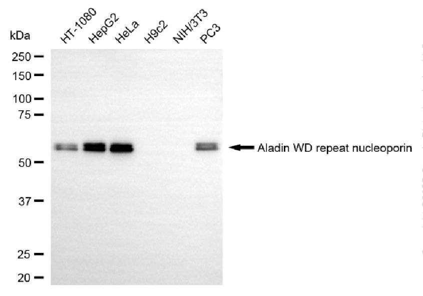
Western blotting analysis using aladin WD repeat nucleoporin antibody. Total cell lysates (30 μg) from various cell lines were loaded and separated by SDS-PAGE. The blot was incubated with aladin WD repeat nucleoporin antibody and HRP-conjugated goat anti-mouse secondary antibody respectively.ICC/IF
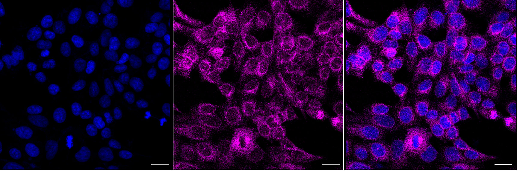
Immunocytochemical staining of HAP1 cells with Aladin WD repeat nucleoporin antibody. Nuclei were stained blue with DAPI; Aladin WD repeat nucleoporin was stained magenta with Alexa Fluor® 647. Images were taken using Leica stellaris 5. Protein abundance based on laser Intensity and smart gain: Medium. Scale bar, 20 μm.FC
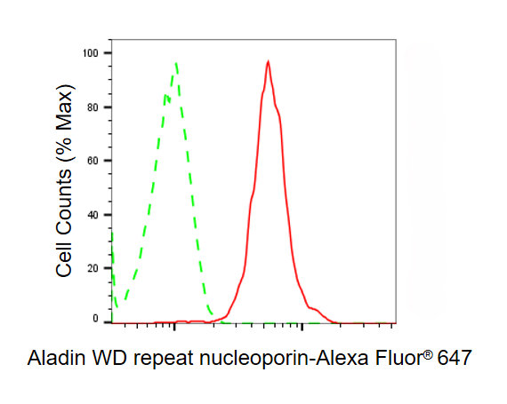
Flow cytometric analysis of Aladin WD repeat nucleoporin expression in HAP-1 cells using Aladin WD repeat nucleoporin antibody. Green, isotype control; red, Aladin WD repeat nucleoporin.| Product Name | Mouse anti-AAAS Monoclonal antibody |
|---|---|
| Antibody Type | Primary Antibodies |
| Immunogen | Recombinant Human AAAS protein |
| Clonality | Monoclonal |
|---|---|
| Isotype | IgG2a |
| Host Species | Mouse |
| Tested Applications | FCICC/IFWB |
| WB:1:500-1:2500 ICC/IF:1:100-1:1000 FC:1:100-1:1000 |
|
| Species Reactivity | Human |
| Concentration | 1mg/ml |
| Purification | Affinity purified |
| Gene Symbol | AAAS |
|---|---|
| Gene Synonyms | AAA AAASb GL003 ALADIN ADRACALA ADRACALIN |
| Gene Full Name | aladin WD repeat nucleoporin |
| Gene Summary | The protein encoded by this gene is a member of the WD-repeat family of regulatory proteins and may be involved in normal development of the peripheral and central nervous system. The encoded protein is part of the nuclear pore complex and is anchored there by NDC1. Defects in this gene are a cause of achalasia-addisonianism-alacrima syndrome (AAAS), also called triple-A syndrome or Allgrove syndrome. Two transcript variants encoding different isoforms have been found for this gene. [provided by RefSeq, Mar 2010] |
| Alternative Names | AAAS Aladin WD Repeat Nucleoporin Adracalin Aladin Achalasia Adrenocortical Insufficiency Alacrimia Allgrove Triple-A ADRACALA Achalasia Adrenocortical Insufficiency Alacrimia (Allgrove Triple-A) AAASb GL003 AAA |
| Cellular Localization | Nucleus, nuclear pore complex. Cytoplasm, cytoskeleton, spindle pole. Nucleus envelope. |
WB
Western blotting analysis using aladin WD repeat nucleoporin antibody. Total cell lysates (30 μg) from various cell lines were loaded and separated by SDS-PAGE. The blot was incubated with aladin WD repeat nucleoporin antibody and HRP-conjugated goat anti-mouse secondary antibody respectively.
ICC/IF
Immunocytochemical staining of HAP1 cells with Aladin WD repeat nucleoporin antibody. Nuclei were stained blue with DAPI; Aladin WD repeat nucleoporin was stained magenta with Alexa Fluor® 647. Images were taken using Leica stellaris 5. Protein abundance based on laser Intensity and smart gain: Medium. Scale bar, 20 μm.
FC
Flow cytometric analysis of Aladin WD repeat nucleoporin expression in HAP-1 cells using Aladin WD repeat nucleoporin antibody. Green, isotype control; red, Aladin WD repeat nucleoporin.| Application Notes | WB:1:500-1:2500 ICC/IF:1:100-1:1000 FC:1:100-1:1000 |
|---|
| Form | Liquid |
|---|---|
| Storage Instructions | Upon receipt, store at -20°C or -80°C. Avoid repeated freeze. |
| Storage Buffer | 0.03% Proclin 300 Constituents: 50% Glycerol, 0.01M PBS, PH 7.4 |


