WB
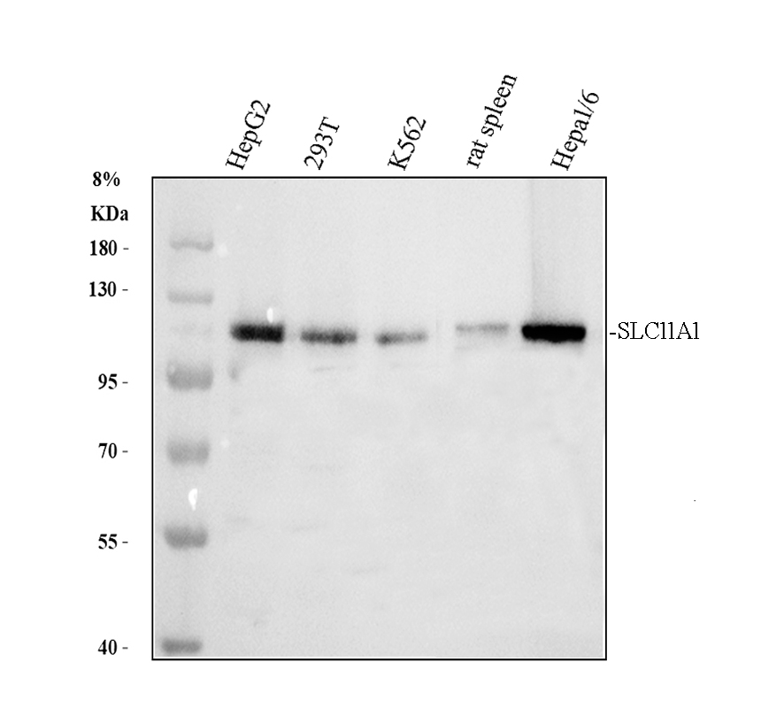
Western blot analysis of SLC11A1 using anti-SLC11A1 antibody. The sample well of each lane was loaded with 30 ug of sample under reducing conditions.After electrophoresis, proteins were transferred to a membrane. Then the membrane was incubated with rabbit anti-SLC11A1 antigen affinity purified polyclonal antibody at a dilution of 1:1000 and probed with a goat anti-rabbit IgG-HRP secondary antibody.The signal is developed using ECL Plus Western Blotting Substrate.IHC
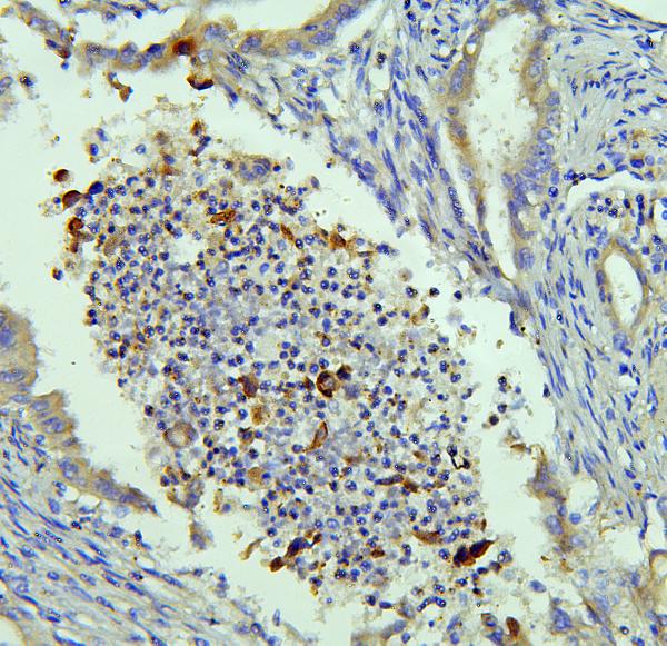
IHC analysis of SLC11A1 using anti-SLC11A1 antibody.SLC11A1 was detected in a paraffin-embedded section of human intestinal cancer tissue. Biotinylated goat anti-rabbit IgG was used as secondary antibody. The tissue section was incubated with rabbit anti-SLC11A1 Antibody at a dilution of 1:200 and developed using Strepavidin-Biotin-Complex (SABC) with DAB as the chromogen.IHC
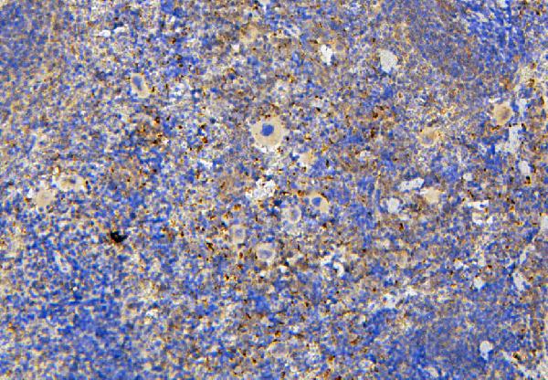
IHC analysis of SLC11A1 using anti-SLC11A1 antibody .SLC11A1 was detected in a paraffin-embedded section of mouse spleen tissue. Biotinylated goat anti-rabbit IgG was used as secondary antibody. The tissue section was incubated with rabbit anti-SLC11A1 Antibody at a dilution of 1:200 and developed using Strepavidin-Biotin-Complex (SABC) with DAB as the chromogen.IHC
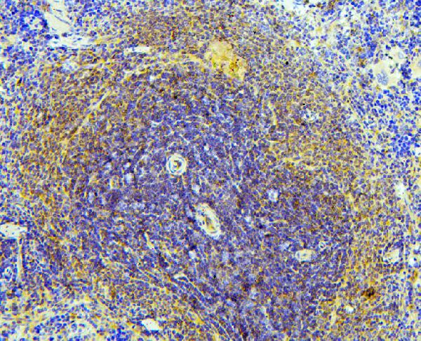
IHC analysis of SLC11A1 using anti-SLC11A1 antibody.SLC11A1 was detected in a paraffin-embedded section of rat spleen tissue. Biotinylated goat anti-rabbit IgG was used as secondary antibody. The tissue section was incubated with rabbit anti-SLC11A1 Antibody at a dilution of 1:200 and developed using Strepavidin-Biotin-Complex (SABC) as the chromogen.ICC/IF
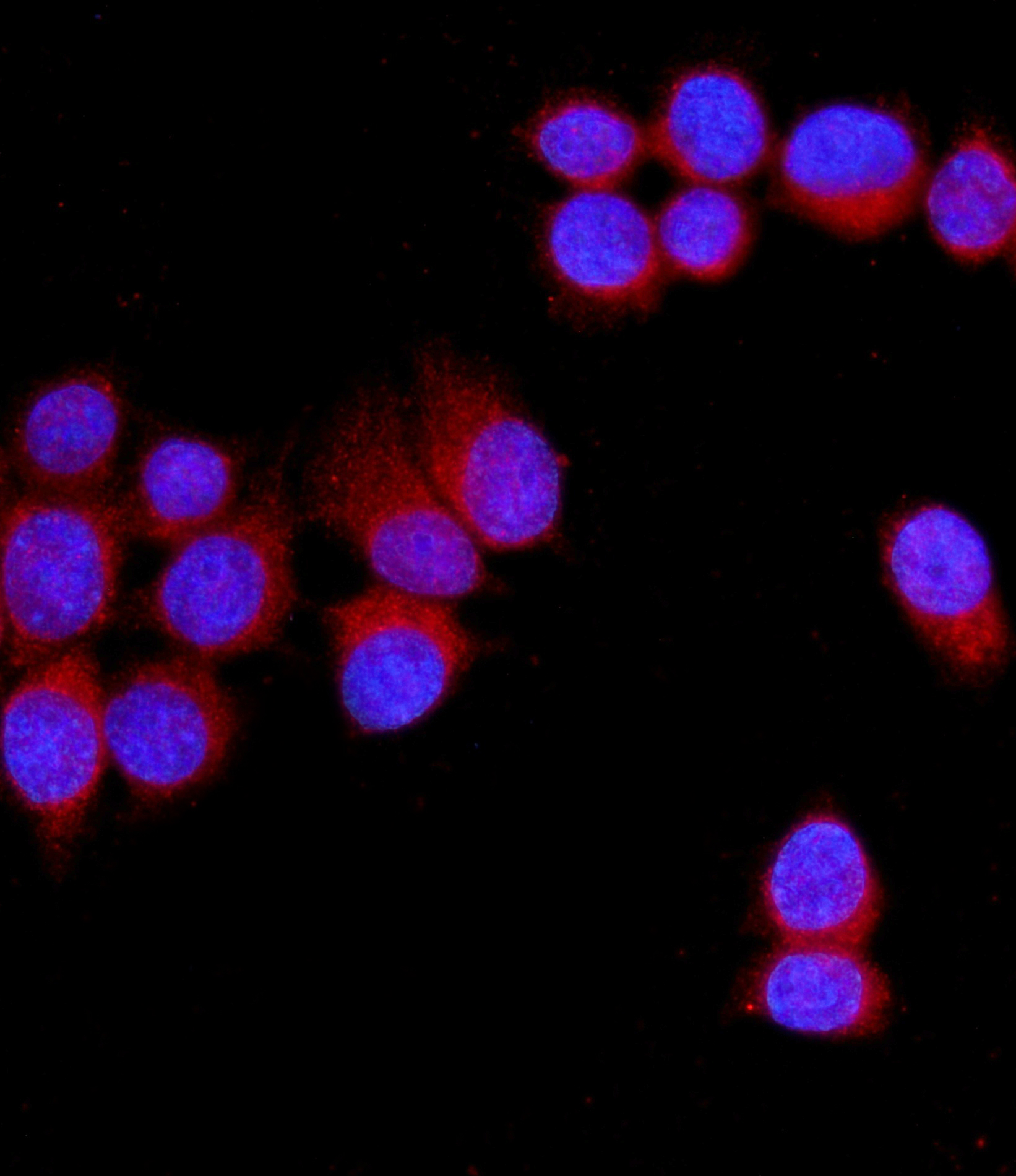
ICC/IF analysis of SLC11A1 using anti-SLC11A1 antibody.SLC11A1 was detected in an immunocytochemical section of MCF-7 cells. The section was incubated with rabbit anti-SLC11A1 Antibody at a dilution of 1:100. Fluoro594-conjugated Anti-rabbit IgG Secondary Antibody (red) was used as secondary antibody. The section was counterstained with DAPI(Blue).| Product Name | Rabbit polyclonal antibody to SLC11A1 |
|---|---|
| Antibody Type | Primary Antibodies |
| Immunogen | E. coli-derived human NRAMP1/SLC11A1 recombinant protein (Position: F183-S549). Human SLC11A1 shares 87.4% and 87.9% amino acid (aa) sequence identity with mouse and rat SLC11A1, respectively. |
| Clonality | Polyclonal |
|---|---|
| Isotype | IgG |
| Host Species | Rabbit |
| Tested Applications | ICC/IFIHCWB |
| WB:1:500-1:2000 ICC/IF:1:50-1:400 IHC:1:50-1:400 |
|
| Species Reactivity | HumanMouseRat |
| Concentration | 0.5mg/ml |
| Purification | Affinity purified |
| Gene Symbol | SLC11A1 |
|---|---|
| Gene Synonyms | LSH NRAMP NRAMP1 |
| Gene Full Name | solute carrier family 11 member 1 |
| Gene Summary | This gene is a member of the solute carrier family 11 (proton-coupled divalent metal ion transporters) family and encodes a multi-pass membrane protein. The protein functions as a divalent transition metal (iron and manganese) transporter involved in iron metabolism and host resistance to certain pathogens. Mutations in this gene have been associated with susceptibility to infectious diseases such as tuberculosis and leprosy, and inflammatory diseases such as rheumatoid arthritis and Crohn disease. Alternatively spliced variants that encode different protein isoforms have been described but the full-length nature of only one has been determined. [provided by RefSeq, Jul 2008] |
| Alternative Names | solute carrier family 11 member 1 LSH NRAMP NRAMP1 SLC11A1 |
| Molecular Weight(MW) | 60 kDa(Observed band size: 90-120 kDa) |
| Cellular Localization | Endosome,Lysosome,Membrane |
WB
Western blot analysis of SLC11A1 using anti-SLC11A1 antibody. The sample well of each lane was loaded with 30 ug of sample under reducing conditions.After electrophoresis, proteins were transferred to a membrane. Then the membrane was incubated with rabbit anti-SLC11A1 antigen affinity purified polyclonal antibody at a dilution of 1:1000 and probed with a goat anti-rabbit IgG-HRP secondary antibody.The signal is developed using ECL Plus Western Blotting Substrate.
IHC
IHC analysis of SLC11A1 using anti-SLC11A1 antibody.SLC11A1 was detected in a paraffin-embedded section of human intestinal cancer tissue. Biotinylated goat anti-rabbit IgG was used as secondary antibody. The tissue section was incubated with rabbit anti-SLC11A1 Antibody at a dilution of 1:200 and developed using Strepavidin-Biotin-Complex (SABC) with DAB as the chromogen.
IHC
IHC analysis of SLC11A1 using anti-SLC11A1 antibody .SLC11A1 was detected in a paraffin-embedded section of mouse spleen tissue. Biotinylated goat anti-rabbit IgG was used as secondary antibody. The tissue section was incubated with rabbit anti-SLC11A1 Antibody at a dilution of 1:200 and developed using Strepavidin-Biotin-Complex (SABC) with DAB as the chromogen.
IHC
IHC analysis of SLC11A1 using anti-SLC11A1 antibody.SLC11A1 was detected in a paraffin-embedded section of rat spleen tissue. Biotinylated goat anti-rabbit IgG was used as secondary antibody. The tissue section was incubated with rabbit anti-SLC11A1 Antibody at a dilution of 1:200 and developed using Strepavidin-Biotin-Complex (SABC) as the chromogen.
ICC/IF
ICC/IF analysis of SLC11A1 using anti-SLC11A1 antibody.SLC11A1 was detected in an immunocytochemical section of MCF-7 cells. The section was incubated with rabbit anti-SLC11A1 Antibody at a dilution of 1:100. Fluoro594-conjugated Anti-rabbit IgG Secondary Antibody (red) was used as secondary antibody. The section was counterstained with DAPI(Blue).| Application Notes | WB:1:500-1:2000 ICC/IF:1:50-1:400 IHC:1:50-1:400 |
|---|
| Form | Liquid |
|---|---|
| Storage Instructions | 12 months from date of receipt, -20℃ as supplied. 6 months 2 to 8℃ after reconstitution. Avoid repeated freezing and thawing. |
| Storage Buffer | 500ug/ml antibody with PBS, 0.02% NaN3, 1 mg/ml BSA and 50% glycerol. |


