WB
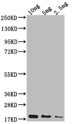
Western blotting analysis using AP2S1 antibody. Total cell lysates (30 μg) from various cell lines were loaded and separated by SDS-PAGE. The blot was incubated with AP2S1 antibody and HRP-conjugated goat anti-rabbit secondary antibody respectively.IHC
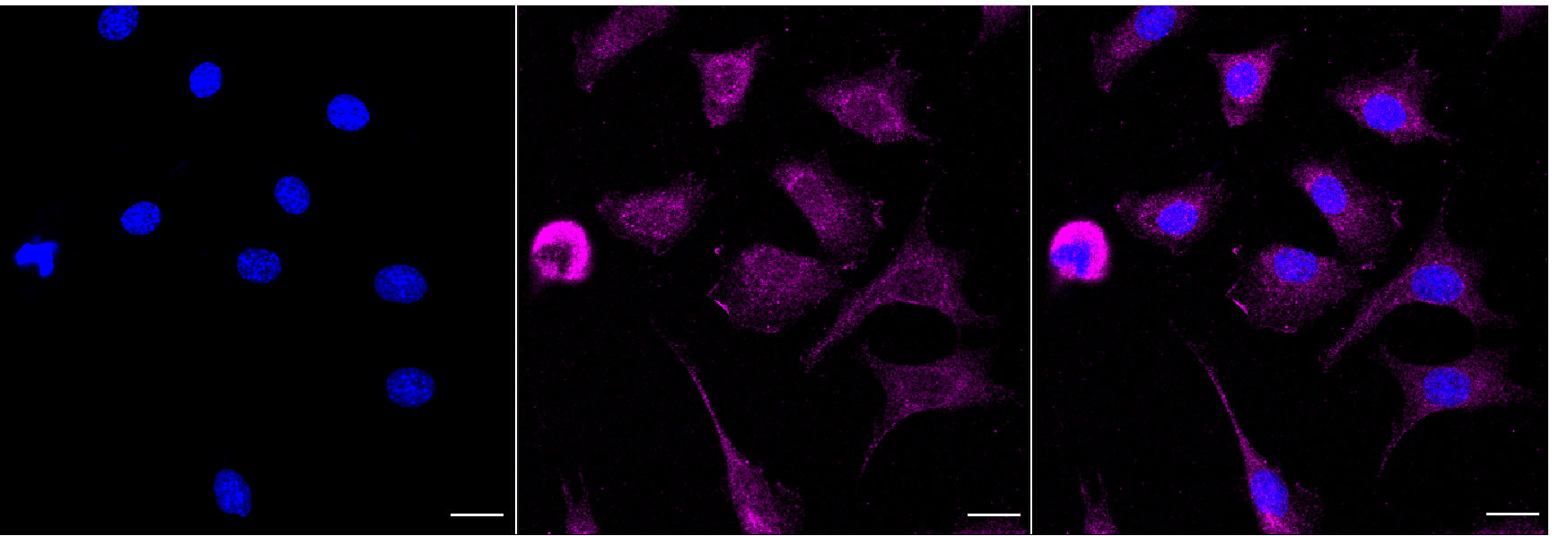
Immunocytochemical staining of C2C12 cells with AP2S1 antibody. Nuclei were stained blue with DAPI; AP2S1 was stained magenta with Alexa Fluor® 647. Images were taken using Leica stellaris 5. Protein abundance based on laser Intensity and smart gain: Medium. Scale bar, 20 μm.FC
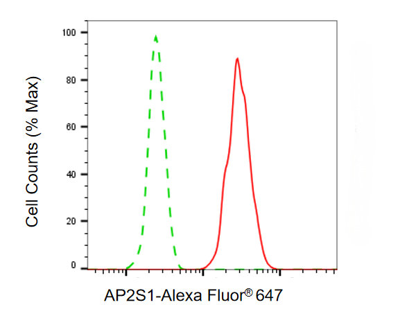
Flow cytometric analysis of AP2S1 expression in C2C12 cells using AP2S1 antibody. Green, isotype control; red, AP2S1.| Product Name | AP2S1 Recombinant Monoclonal Antibody |
|---|---|
| Antibody Type | Primary Antibodies |
| Immunogen | A synthesized peptide derived from Human AP2S1 protein |
| Clonality | Monoclonal |
|---|---|
| Isotype | IgG |
| Host Species | Rabbit |
| Tested Applications | FCICC/IFWB |
| WB:1:1000-1:5000 FC:1:200-1:2000 ICC/IF:1:100-1:1000 |
|
| Species Reactivity | HumanMouseRat |
| Concentration | 1mg/ml |
| Purification | Affinity purified |
| Gene Symbol | AP2S1 |
|---|---|
| Gene Synonyms | AP17 FBH3 HHC3 FBHOk CLAPS2 |
| Gene Full Name | adaptor related protein complex 2 subunit sigma 1 |
| Gene Summary | One of two major clathrin-associated adaptor complexes, AP-2, is a heterotetramer which is associated with the plasma membrane. This complex is composed of two large chains, a medium chain, and a small chain. This gene encodes the small chain of this complex. Alternative splicing results in multiple transcript variants. [provided by RefSeq, Jul 2014] |
| Alternative Names | AP2S1 Adaptor Related Protein Complex 2 Subunit Sigma 1 CLAPS2 FBH3 Adaptor Related Protein Complex 2 Sigma 1 Subunit Clathrin Assembly Protein 2 Sigma Small Chain Plasma Membrane Adaptor AP-2 17 KDa Protein Adaptor Protein Complex AP-2 Subunit Sigma Clathrin Coat-Associated Protein AP17 Clathrin Coat Assembly Protein AP17 AP-2 Complex Subunit Sigma HA2 17 KDa Subunit Sigma2-Adaptin Sigma-2 FBHOk AP17 HHC3 Clathrin-Associated/Assembly/Adaptor Protein Small 2 (17kD) Adaptor-Related Protein Complex 2 Subunit Sigma Hypocalciuric Hypercalcemia 3 (Oklahoma Type) FBHOK |
| Cellular Localization | Cell membrane. Membrane, coated pit; Peripheral membrane protein; Cytoplasmic side. |
WB
Western blotting analysis using AP2S1 antibody. Total cell lysates (30 μg) from various cell lines were loaded and separated by SDS-PAGE. The blot was incubated with AP2S1 antibody and HRP-conjugated goat anti-rabbit secondary antibody respectively.
IHC
Immunocytochemical staining of C2C12 cells with AP2S1 antibody. Nuclei were stained blue with DAPI; AP2S1 was stained magenta with Alexa Fluor® 647. Images were taken using Leica stellaris 5. Protein abundance based on laser Intensity and smart gain: Medium. Scale bar, 20 μm.
FC
Flow cytometric analysis of AP2S1 expression in C2C12 cells using AP2S1 antibody. Green, isotype control; red, AP2S1.| Application Notes | WB:1:1000-1:5000 FC:1:200-1:2000 ICC/IF:1:100-1:1000 |
|---|
| Form | Liquid |
|---|---|
| Storage Instructions | Upon receipt, store at -20°C or -80°C. Avoid repeated freeze. |
| Storage Buffer | PBS (pH 7.4) containing 50% glycerol, and 0.02% sodium azide. |


