Application
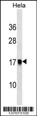
AP2S1 Antibody (Center) western blot analysis in Hela cell line lysates (35ug/lane).This demonstrates the AP2S1 antibody detected the AP2S1 protein (arrow).| Product Name | AP2S1 Antibody (Center) |
|---|---|
| Antibody Type | Primary Antibodies |
| Antigen Alias | AP2S1; AP17; CLAPS2; AP-2 complex subunit sigma; Adapter-related protein complex 2 sigma subunit; Adaptor protein complex AP-2 subunit sigma; Clathrin assembly protein 2 small chain; Clathrin coat assembly protein AP17; Clathrin coat-associated protein AP1 |
| Clonality | Polyclonal |
|---|---|
| Isotype | Ig |
| Host Species | Rabbit |
| Tested Applications | WB |
| WB:1:100~500 |
|
| Species Reactivity | Human |
| Concentration | 1mg/ml |
| Gene Synonyms | AP17 CLAPS2 |
|---|---|
| Alternative Names | AP2S1 AP17 CLAPS2 AP-2 complex subunit sigma Adapter-related protein complex 2 sigma subunit Adaptor protein complex AP-2 subunit sigma Clathrin assembly protein 2 small chain Clathrin coat assembly protein AP17 Clathrin coat-associated protein AP1 |
| Molecular Weight(MW) | 17018 Da |
| Function | Component of the adaptor protein complex 2 (AP-2). Adaptor protein complexes function in protein Transport via Transport vesicles in different membrane traffic pathways. Adaptor protein complexes are vesicle coat components and appear to be involved in cargo selection and vesicle formation. AP-2 is involved in clathrin-dependent endocytosis in which cargo proteins are incorporated into vesicles surrounded by clathrin (clathrin- coated vesicles, CCVs) which are destined for fusion with the early endosome. The clathrin lattice serves as a mechanical scaffold but is itself unable to bind directly to membrane components. Clathrin-associated adaptor protein (AP) complexes which can bind directly to both the clathrin lattice and to the lipid and protein components of membranes are considered to be the major clathrin adaptors contributing the CCV formation. AP-2 also serves as a cargo receptor to selectively sort the membrane proteins involved in receptor-mediated endocytosis. AP-2 seems to play a role in the recycling of synaptic vesicle membranes from the presynaptic surface. AP-2 recognizes Y-X-X-[FILMV] (Y-X-X-Phi) and [ED]-X-X-X-L-[LI] endocytosis signal motifs within the cytosolic tails of transmembrane cargo molecules. AP-2 may also play a role in maintaining normal post-endocytic trafficking through the ARF6-regulated, non-clathrin pathway. The AP-2 alpha and AP-2 sigma subunits are thought to contribute to the recognition of the [ED]-X-X-X-L-[LI] motif (By similarity) |
| Tissue Specificity | This AP2S1 antibody is generated from rabbits immunized with a KLH conjugated synthetic peptide between 35-64 amino acids from the Central region of human AP2S1. |
| Cellular Localization | Cell membrane. Membrane, coated pit; Peripheral membrane protein; Cytoplasmic side. Note=AP-2 appears to be excluded from internalizing CCVs and to disengage from sites of endocytosis seconds before internalization of the nascent CCV (By similarity) |
| Entrez Gene | 1175 |
|---|---|
| Nucleotide Accession | NP_004060.2 NP_067586.1 |
Application
AP2S1 Antibody (Center) western blot analysis in Hela cell line lysates (35ug/lane).This demonstrates the AP2S1 antibody detected the AP2S1 protein (arrow).| Application Notes | WB:1:100~500 |
|---|
| Form | Liquid |
|---|---|
| Storage Instructions | For short-term storage, store at 4° C. For long-term storage, aliquot and store at -20ºC or below. Avoid multiple freeze-thaw cycles. |
| Storage Buffer | Purified polyclonal antibody supplied in PBS with 0.09% (W/V) sodium azide. This antibody is purified through a protein A column, followed by peptide affinity purification. |


