WB
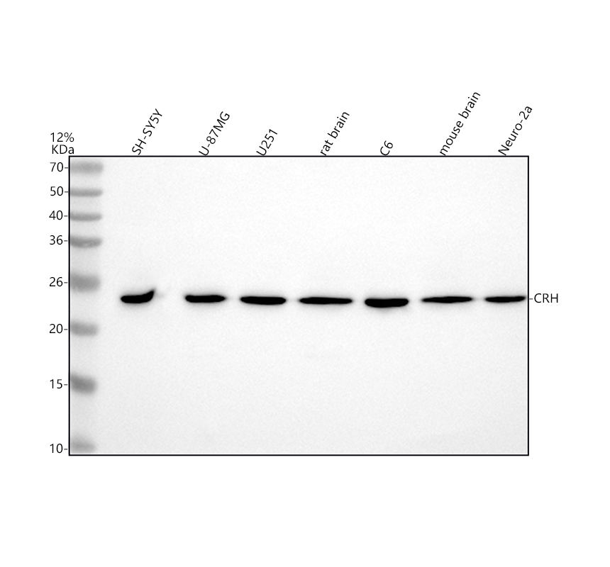
Western blot analysis of CRF/CRH using anti-CRF/CRH antibody. The sample well of each lane was loaded with 30 ug of sample under reducing conditions.After electrophoresis, proteins were transferred to a membrane. Then the membrane was incubated with rabbit anti-CRF/CRH antigen affinity purified polyclonal antibody at a dilution of 1:1000 and probed with a goat anti-rabbit IgG-HRP secondary antibody. The signal is developed using ECL Plus Western Blotting Substrate.FC
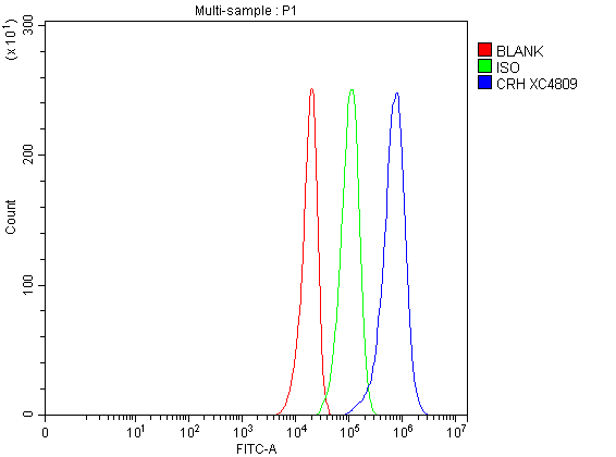
Flow Cytometry analysis of U251 cells using anti-CRF/CRH antibody.Overlay histogram showing U251 cells stained with anti-CRF/CRH antibody (Blue line). The cells were fixed with 4% paraformaldehyde and blocked with 10% normal goat serum. And then incubated with rabbit anti-CRF/CRH Antibody at 1:100 dilution for 30 min at 20°C. Fluoro488 conjugated goat anti-rabbit IgG was used as secondary antibody at 1:100 dilution for 30 minutes at 20°C. Isotype control antibody (Green line) was rabbit IgG at 1:100 dilution used under the same conditions. Unlabelled sample without incubation with primary antibody and secondary antibody (Red line) was used as a blank control.| Product Name | Rabbit polyclonal antibody to CRF/CRH |
|---|---|
| Antibody Type | Primary Antibodies |
| Immunogen | A synthetic peptide corresponding to a sequence at the C-terminus of human CRF/CRH, identical to the related mouse and rat sequences. |
| Clonality | Polyclonal |
|---|---|
| Isotype | IgG |
| Host Species | Rabbit |
| Tested Applications | FCWB |
| WB:1:500-1:2000 FC:1:50-1:200 |
|
| Species Reactivity | HumanMouseRat |
| Concentration | 1mg/ml |
| Purification | Affinity purified |
| Gene Symbol | CRH |
|---|---|
| Gene Synonyms | CRF CRH1 |
| Gene Full Name | corticotropin releasing hormone |
| Gene Summary | This gene encodes a member of the corticotropin-releasing factor family. The encoded preproprotein is proteolytically processed to generate the mature neuropeptide hormone. In response to stress, this hormone is secreted by the paraventricular nucleus (PVN) of the hypothalamus, binds to corticotropin releasing hormone receptors and stimulates the release of adrenocorticotropic hormone from the pituitary gland. Marked reduction in this protein has been observed in association with Alzheimer's disease. Autosomal recessive hypothalamic corticotropin deficiency has multiple and potentially fatal metabolic consequences including hypoglycemia and hepatitis. In addition to production in the hypothalamus, this protein is also synthesized in peripheral tissues, such as T lymphocytes, and is highly expressed in the placenta. In the placenta it is a marker that determines the length of gestation and the timing of parturition and delivery. A rapid increase in circulating levels of the hormone occurs at the onset of parturition, suggesting that, in addition to its metabolic functions, this protein may act as a trigger for parturition. [provided by RefSeq, Nov 2015] |
| Alternative Names | CRH CRF Corticotropin-releasing hormone Corticotropin-releasing factor Corticotropin releasing factor |
| Molecular Weight(MW) | 21 kDa(Observed band size: 25 kDa) |
| Cellular Localization | Secreted |
WB
Western blot analysis of CRF/CRH using anti-CRF/CRH antibody. The sample well of each lane was loaded with 30 ug of sample under reducing conditions.After electrophoresis, proteins were transferred to a membrane. Then the membrane was incubated with rabbit anti-CRF/CRH antigen affinity purified polyclonal antibody at a dilution of 1:1000 and probed with a goat anti-rabbit IgG-HRP secondary antibody. The signal is developed using ECL Plus Western Blotting Substrate.
FC
Flow Cytometry analysis of U251 cells using anti-CRF/CRH antibody.Overlay histogram showing U251 cells stained with anti-CRF/CRH antibody (Blue line). The cells were fixed with 4% paraformaldehyde and blocked with 10% normal goat serum. And then incubated with rabbit anti-CRF/CRH Antibody at 1:100 dilution for 30 min at 20°C. Fluoro488 conjugated goat anti-rabbit IgG was used as secondary antibody at 1:100 dilution for 30 minutes at 20°C. Isotype control antibody (Green line) was rabbit IgG at 1:100 dilution used under the same conditions. Unlabelled sample without incubation with primary antibody and secondary antibody (Red line) was used as a blank control.| Application Notes | WB:1:500-1:2000 FC:1:50-1:200 |
|---|
| Form | Liquid |
|---|---|
| Storage Instructions | 12 months from date of receipt,-20℃ as supplied. |
| Storage Buffer | 500 ug/ml antibody with PBS, 0.02% NaN3, 1 mg/ml BSA and 50% glycerol. |


