WB
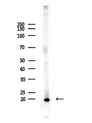
Western blot analysis of lysates from A375 cells(heat-shock,45min treatment), using SCXA Antibody at 1/1000 dilution. 5ug/NC membrane strip. Exposure for 30s with ECL Kit. Bands result from membrane strip incubation.WB
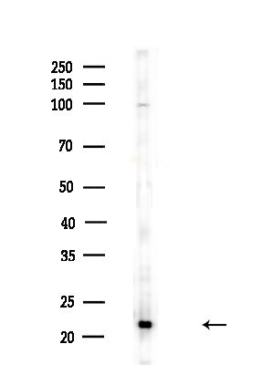
Western blot analysis of lysates from Rat lung, using SCXA Antibody at 1/1000 dilution. 5ug/NC membrane strip. Exposure for 30s with ECL Kit. Bands result from membrane strip incubation.WB
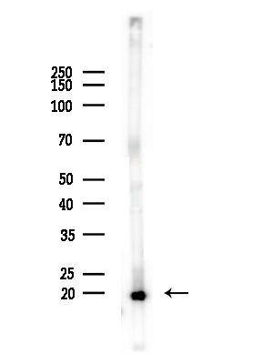
Western blot analysis of lysates from Mouse brain, using SCXA Antibody at 1/1000 dilution. 5ug/NC membrane strip. Exposure for 30s with ECL Kit. Bands result from membrane strip incubation.IHC
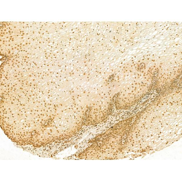
Rabbit polyclonal antibody to SCXAat 1/100 staining Human esophageal cancer and adjacent normal tissues by IHC-P. The sample was formaldehyde fixed and a heat mediated antigen retrieval step in citrate buffer was performed. The sample was then blocked and incubated with the primary antibody at 4°C overnight. An HRP conjugated anti-Rabbit antibody was used as the secondary antibody.IHC
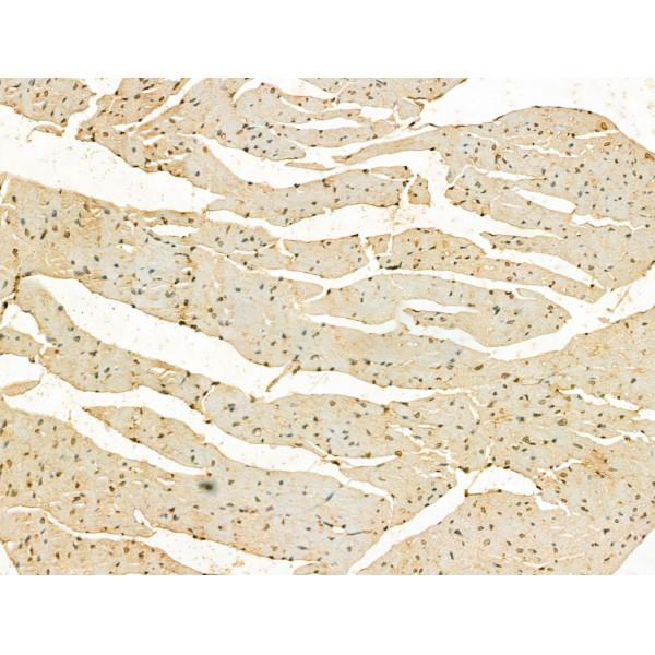
Rabbit polyclonal antibody to SCXA at 1/100 staining Mouse heart tissue by IHC-P. The sample was formaldehyde fixed and a heat mediated antigen retrieval step in citrate buffer was performed. The sample was then blocked and incubated with the primary antibody at 4°C overnight. An HRP conjugated anti-Rabbit antibody was used as the secondary antibody.IHC
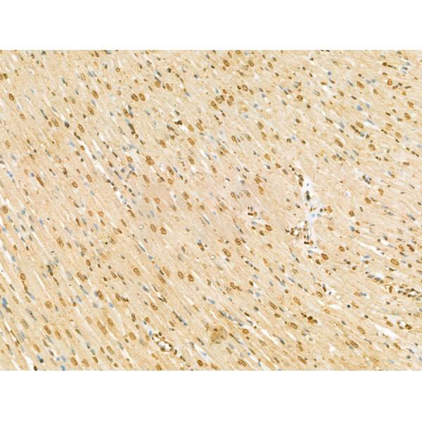
Rabbit polyclonal antibody to SCXA at 1/100 staining Rat heart tissue by IHC-P. The sample was formaldehyde fixed and a heat mediated antigen retrieval step in citrate buffer was performed. The sample was then blocked and incubated with the primary antibody at 4°C overnight. An HRP conjugated anti-Rabbit antibody was used as the secondary antibody.| Product Name | Rabbit polyclonal antibody to SCXA |
|---|---|
| Antibody Type | Primary Antibodies |
| Immunogen | A synthesized peptide derived from human SCXA, corresponding to a region within the internal amino acids. |
| Clonality | Polyclonal |
|---|---|
| Isotype | IgG |
| Host Species | Rabbit |
| Tested Applications | IHCWB |
| WB:1:500-1:2000 IHC:1:50-1:200 |
|
| Species Reactivity | HumanMouseRat |
| Concentration | 1mg/ml |
| Purification | Affinity purified |
| Gene Symbol | SCX |
|---|---|
| Gene Synonyms | SCXA SCXB bHLHa48 |
| Gene Full Name | scleraxis bHLH transcription factor |
| Gene Summary | Enables DNA-binding transcription activator activity, RNA polymerase II-specific and bHLH transcription factor binding activity. Contributes to E-box binding activity. Involved in positive regulation of transcription by RNA polymerase II. Located in nucleus. Part of transcription regulator complex. [provided by Alliance of Genome Resources, Jul 2025] |
| Alternative Names | Basic helix loop helix transcription factor scleraxis Basic helix-loop-helix transcription factor scleraxis bHLHa41 bHLHa48 Class A basic helix-loop-helix protein 41 Class A basic helix-loop-helix protein 48 scleraxis scleraxis homolog A SCX SCX_HUMAN SCXB |
| Molecular Weight(MW) | 22 kDa(Observed band size: 21 kDa) |
| Cellular Localization | Nucleus |
WB
Western blot analysis of lysates from A375 cells(heat-shock,45min treatment), using SCXA Antibody at 1/1000 dilution. 5ug/NC membrane strip. Exposure for 30s with ECL Kit. Bands result from membrane strip incubation.
WB
Western blot analysis of lysates from Rat lung, using SCXA Antibody at 1/1000 dilution. 5ug/NC membrane strip. Exposure for 30s with ECL Kit. Bands result from membrane strip incubation.
WB
Western blot analysis of lysates from Mouse brain, using SCXA Antibody at 1/1000 dilution. 5ug/NC membrane strip. Exposure for 30s with ECL Kit. Bands result from membrane strip incubation.
IHC
Rabbit polyclonal antibody to SCXAat 1/100 staining Human esophageal cancer and adjacent normal tissues by IHC-P. The sample was formaldehyde fixed and a heat mediated antigen retrieval step in citrate buffer was performed. The sample was then blocked and incubated with the primary antibody at 4°C overnight. An HRP conjugated anti-Rabbit antibody was used as the secondary antibody.
IHC
Rabbit polyclonal antibody to SCXA at 1/100 staining Mouse heart tissue by IHC-P. The sample was formaldehyde fixed and a heat mediated antigen retrieval step in citrate buffer was performed. The sample was then blocked and incubated with the primary antibody at 4°C overnight. An HRP conjugated anti-Rabbit antibody was used as the secondary antibody.
IHC
Rabbit polyclonal antibody to SCXA at 1/100 staining Rat heart tissue by IHC-P. The sample was formaldehyde fixed and a heat mediated antigen retrieval step in citrate buffer was performed. The sample was then blocked and incubated with the primary antibody at 4°C overnight. An HRP conjugated anti-Rabbit antibody was used as the secondary antibody.| Application Notes | WB:1:500-1:2000 IHC:1:50-1:200 |
|---|
| Form | Liquid |
|---|---|
| Storage Instructions | Store at -20 °C. Stable for 12 months from date of receipt. |
| Storage Buffer | Rabbit IgG in phosphate buffered saline , pH 7.4, 150mM NaCl, 0.02% sodium azide and 50% glycerol. |


