WB
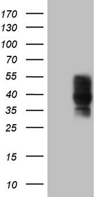
HEK293T cells were transfected with the pCMV6-ENTRY control (Left lane) or pCMV6-ENTRY PDCD1 (Right lane) cDNA for 48 hrs and lysed. Equivalent amounts of cell lysates (5 ug per lane) were separated by SDS-PAGE and immunoblotted with anti-PDCD1.IHC
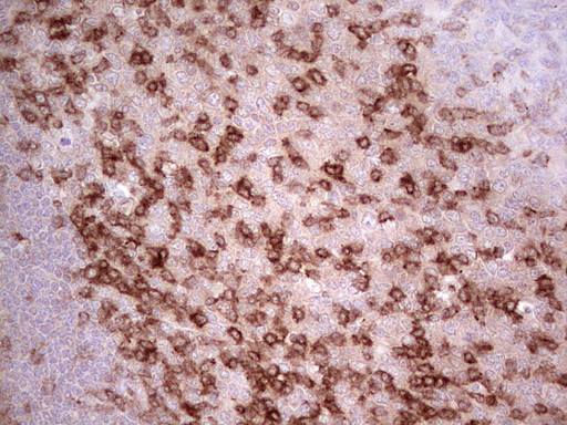
Immunohistochemical staining of paraffin-embedded Human tonsil using anti-PDCD1 mouse monoclonal antibody. Heat-induced epitope retrieval by EDTA solution buffer pH 8.0 at 120°C for 3 min.ICC/IF
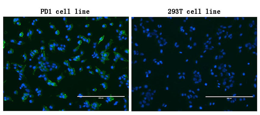
Immunofluorescent staining of PDCD1 stable-transfected HEK293T cells (left) labeling PDCD1 with mouse monoclonal antibody (green) and nucleus with Hoechst33342 (blue). HEK293T cells serve as negative control (right) (1:100).FC
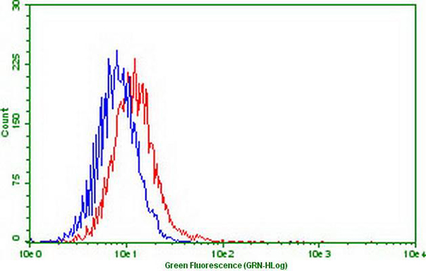
Flow cytometric Analysis of stable expression PD1 cells using anti-PDCD1 antibody (Red) compared to a nonspecific negative control antibody (Blue) (1:50).| Product Name | PD1 (PDCD1) Mouse Monoclonal Antibody |
|---|---|
| Antibody Type | Primary Antibodies |
| Immunogen | Full length human recombinant protein of human PDCD1 (NP_005009) produced in HEK293T cell. |
| Clonality | monoclonal |
|---|---|
| Isotype | IgG2a |
| Host Species | Mouse |
| Tested Applications | FCICC/IFIHCWB |
| WB:1:2000 IHC:1:150 ICC/IF:1:100-1:900 FC:1:50 |
|
| Species Reactivity | Human |
| Concentration | 1mg/ml |
| Purification | Protein A/G |
| Gene Symbol | PDCD1 |
|---|---|
| Gene Synonyms | PD1 PD-1 CD279 SLEB2 hPD-1 hPD-l hSLE1 ADMIO4 AIMTBS |
| Gene Full Name | programmed cell death 1 |
| Gene Summary | Programmed cell death protein 1 (PDCD1) is an immune-inhibitory receptor expressed in activated T cells; it is involved in the regulation of T-cell functions, including those of effector CD8+ T cells. In addition, this protein can also promote the differentiation of CD4+ T cells into T regulatory cells. PDCD1 is expressed in many types of tumors including melanomas, and has demonstrated to play a role in anti-tumor immunity. Moreover, this protein has been shown to be involved in safeguarding against autoimmunity, however, it can also contribute to the inhibition of effective anti-tumor and anti-microbial immunity. [provided by RefSeq, Aug 2020] |
| Alternative Names | CD279 hPD-1 hPD-l hSLE1 PD-1 PD1 SLEB2 |
| Molecular Weight(MW) | 29.2 kDa |
| Cellular Localization | Cell membrane,Membrane. |
WB
HEK293T cells were transfected with the pCMV6-ENTRY control (Left lane) or pCMV6-ENTRY PDCD1 (Right lane) cDNA for 48 hrs and lysed. Equivalent amounts of cell lysates (5 ug per lane) were separated by SDS-PAGE and immunoblotted with anti-PDCD1.
IHC
Immunohistochemical staining of paraffin-embedded Human tonsil using anti-PDCD1 mouse monoclonal antibody. Heat-induced epitope retrieval by EDTA solution buffer pH 8.0 at 120°C for 3 min.
ICC/IF
Immunofluorescent staining of PDCD1 stable-transfected HEK293T cells (left) labeling PDCD1 with mouse monoclonal antibody (green) and nucleus with Hoechst33342 (blue). HEK293T cells serve as negative control (right) (1:100).
FC
Flow cytometric Analysis of stable expression PD1 cells using anti-PDCD1 antibody (Red) compared to a nonspecific negative control antibody (Blue) (1:50).| Application Notes | WB:1:2000 IHC:1:150 ICC/IF:1:100-1:900 FC:1:50 |
|---|
| Form | Liquid |
|---|---|
| Storage Instructions | Store at -20°C as received. |
| Storage Buffer | PBS (pH 7.3) containing 1% BSA, 50% glycerol and 0.02% sodium azide |


