WB
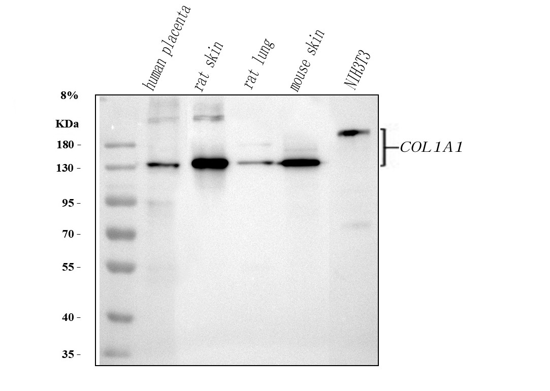
Western blot analysis of anti-Collagen Type I/COL1A1 antibody. The sample well of each lane was loaded with 30 ug of sample under reducing conditions. After electrophoresis, proteins were transferred to a membrane. Then the membrane was incubated with rabbit anti-Collagen Type I/COL1A1 antigen affinity purified polyclonal antibody and probed with a goat anti-rabbit IgG-HRP secondary antibody. The signal is developed using ECL Plus Western Blotting Substrate.IHC
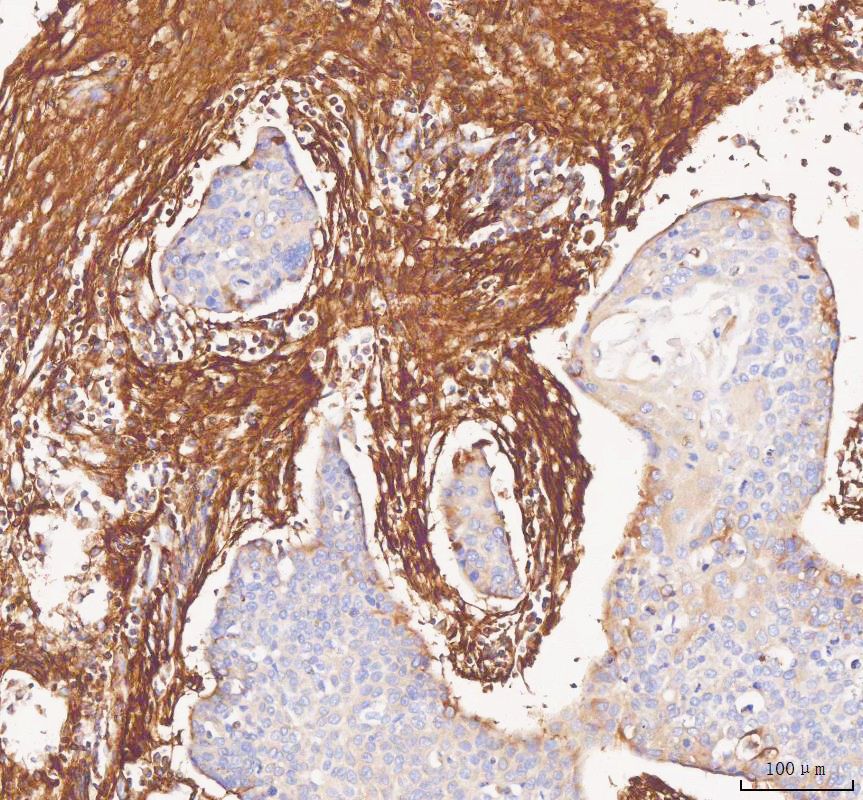
IHC analysis of Collagen Type I/COL1A1 using anti-Collagen Type I/COL1A1 antibody. Collagen Type I/COL1A1 was detected in a paraffin-embedded section of human liver cancer tissue. The tissue section was developed using HRP Conjugated Rabbit IgG Super Vision Assay Kit with DAB as the chromogen.IHC
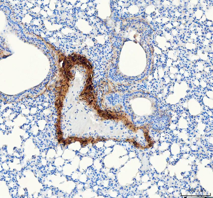
IHC analysis of Collagen Type I/COL1A1 using anti-Collagen Type I/COL1A1 antibody. Collagen Type I/COL1A1 was detected in a paraffin-embedded section of rat lung tissue. The tissue section was developed using HRP Conjugated Rabbit IgG Super Vision Assay Kit with DAB as the chromogen.IHC
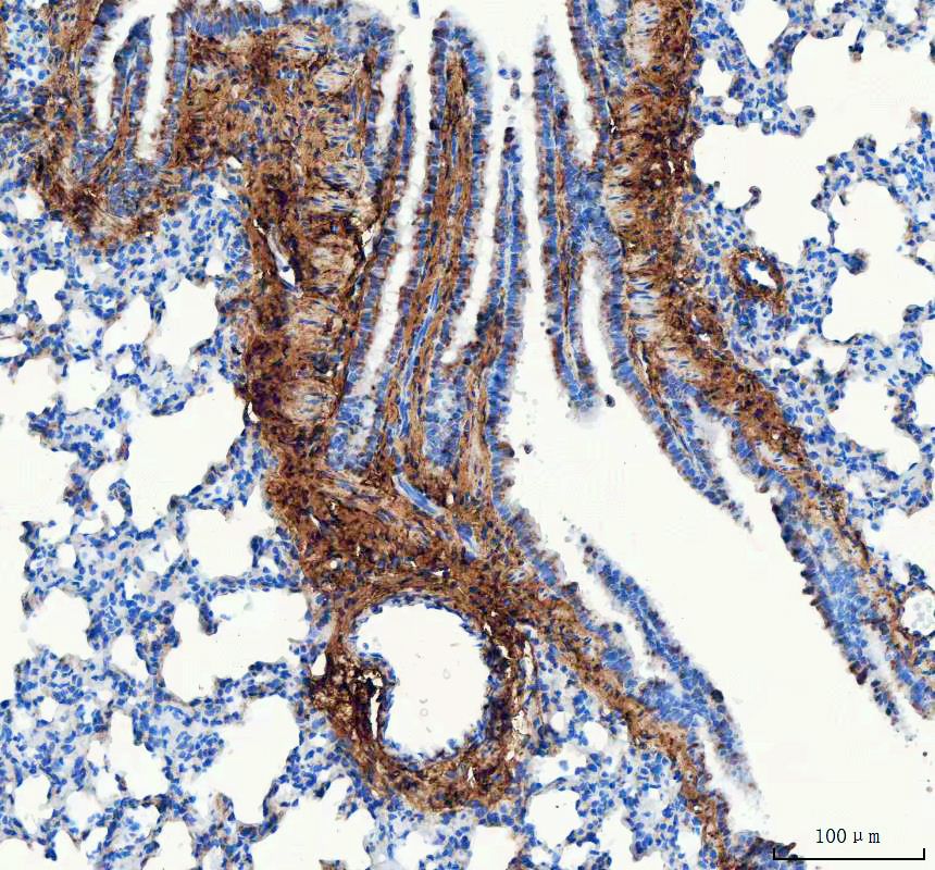
IHC analysis of Collagen Type I/COL1A1 using anti-Collagen Type I/COL1A1 antibody. Collagen Type I/COL1A1 was detected in a paraffin-embedded section of mouse lung tissue. The tissue section was developed using HRP Conjugated Rabbit IgG Super Vision Assay Kit with DAB as the chromogen.IF-P
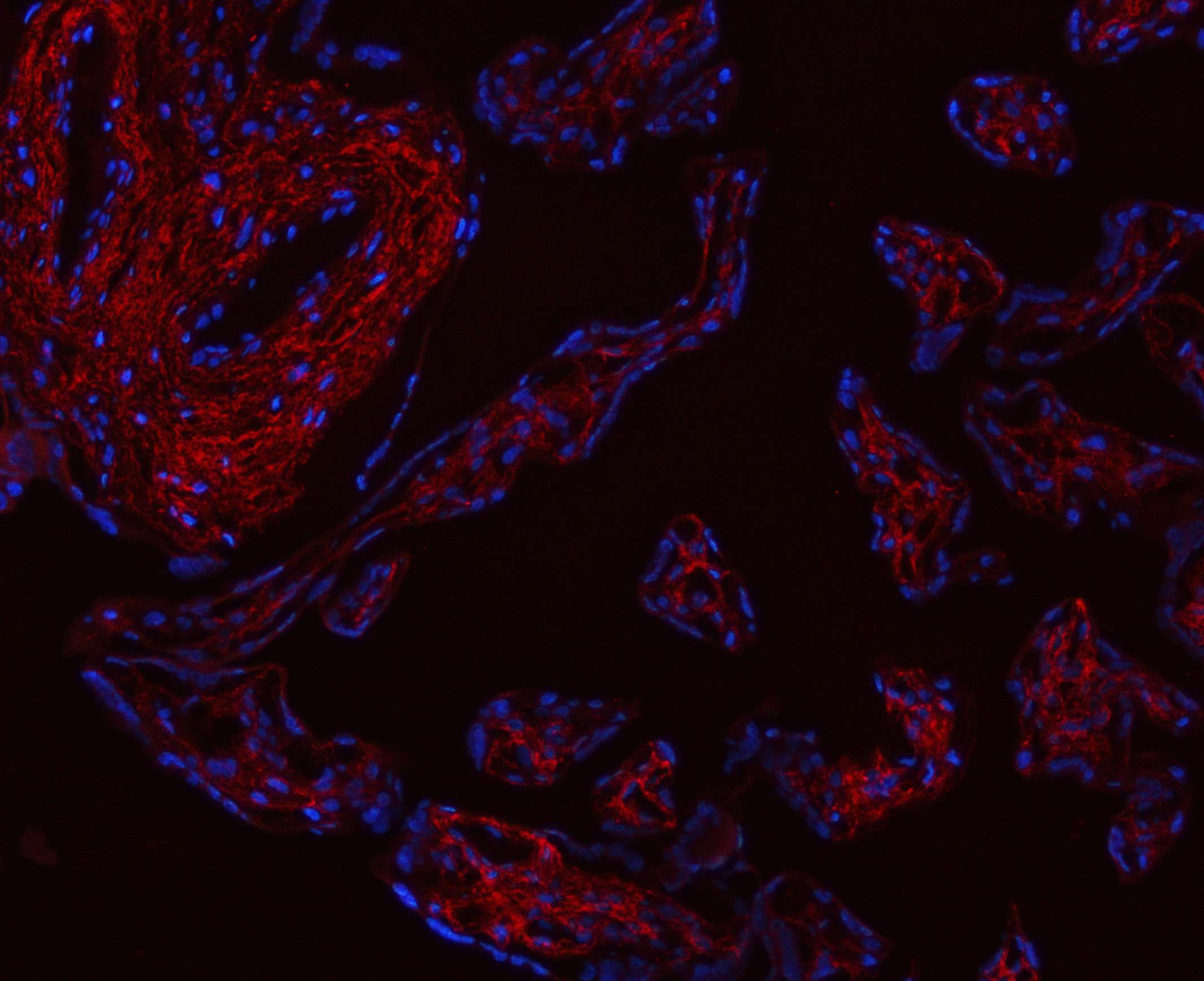
IF analysis of Collagen Type I/COL1A1 using anti-Collagen Type I/COL1A1 antibody. Collagen Type I/COL1A1 was detected in a paraffin-embedded section of human placenta tissue. Cy3-conjugated Anti-rabbit IgG Secondary Antibody (red) was used as secondary antibody. The section was counterstained with DAPI (Blue).IF-P
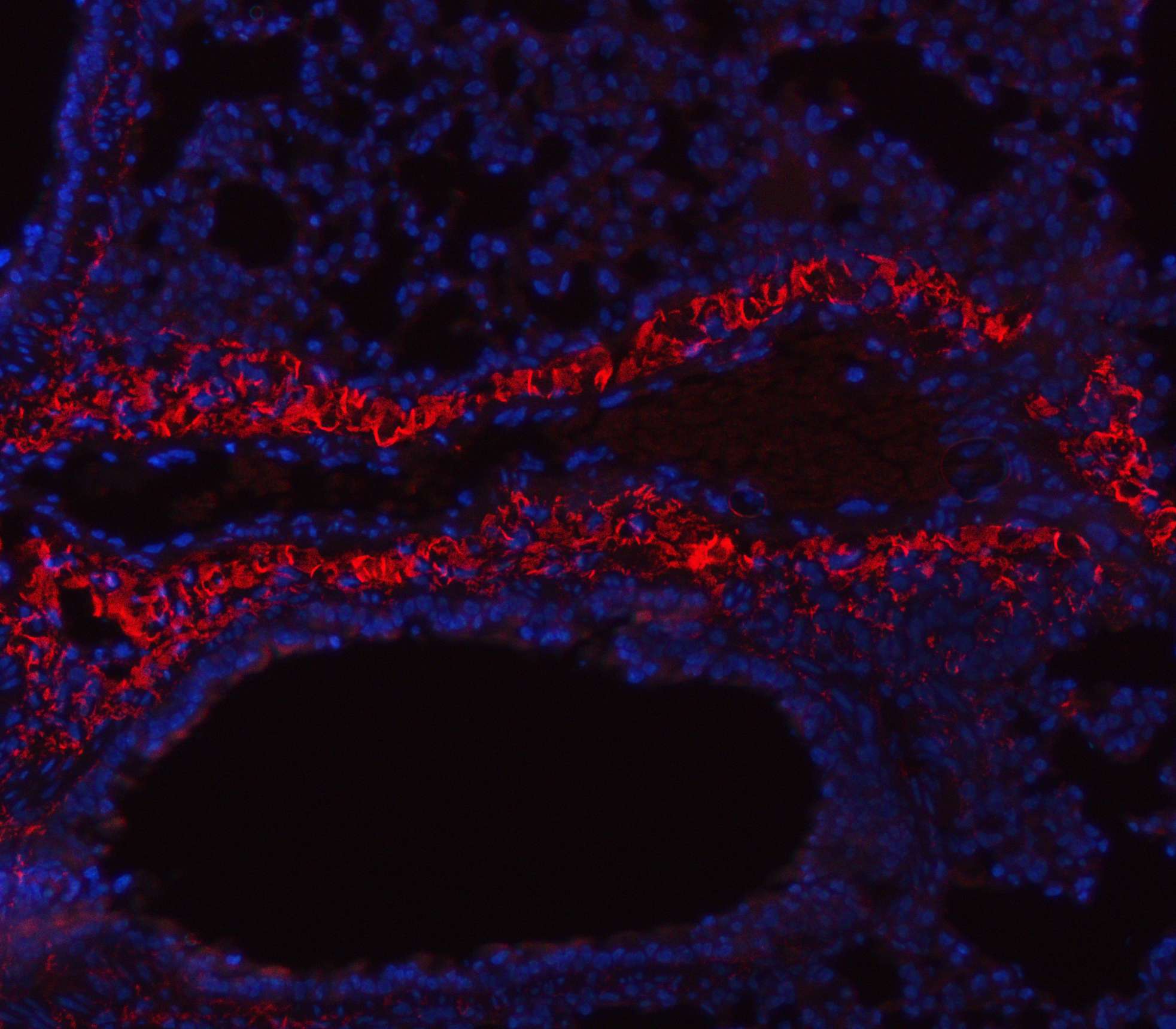
IF analysis of Collagen Type I/COL1A1 using anti-Collagen Type I/COL1A1 antibody. Collagen Type I/COL1A1 was detected in a paraffin-embedded section of mouse lung tissue. Cy3-conjugated Anti-rabbit IgG Secondary Antibody (red) was used as secondary antibody. The section was counterstained with DAPI (Blue).IF-P
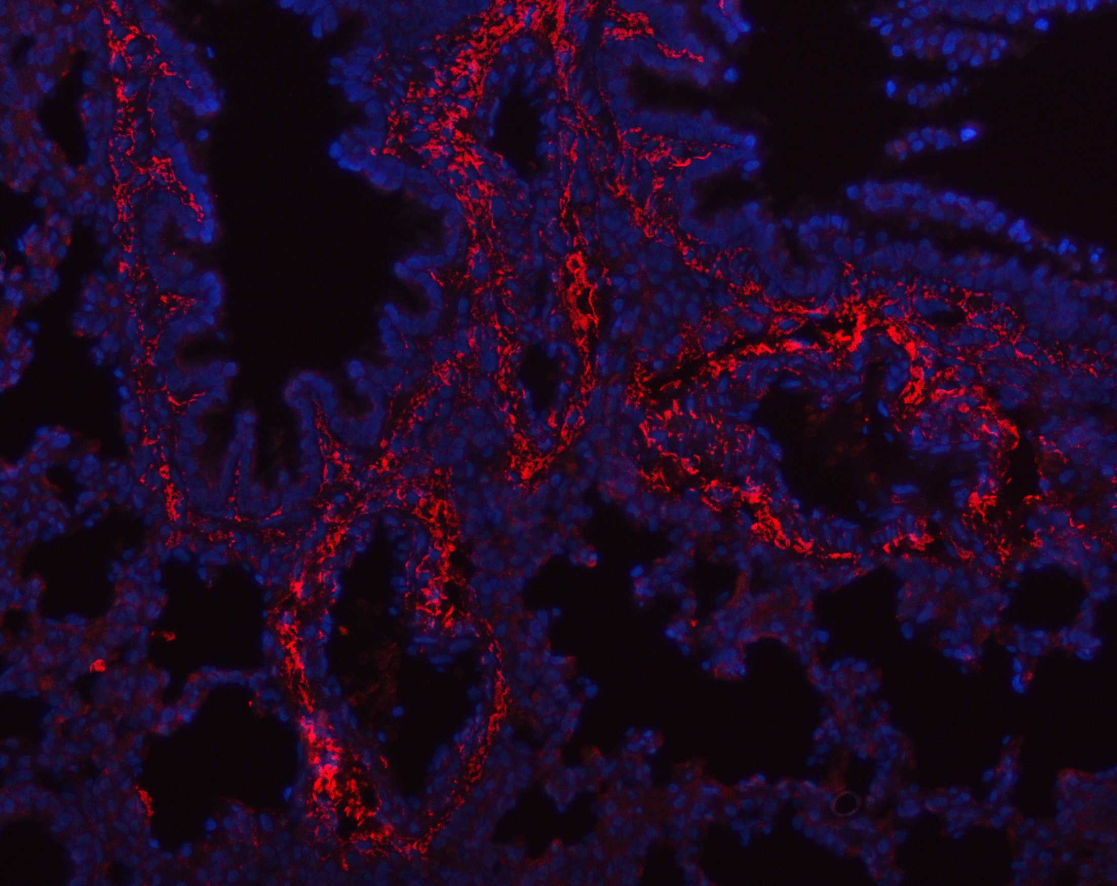
IF analysis of Collagen Type I/COL1A1 using anti-Collagen Type I/COL1A1 antibody. Collagen Type I/COL1A1 was detected in a paraffin-embedded section of rat lung tissue. Cy3-conjugated Anti-rabbit IgG Secondary Antibody (red) was used as secondary antibody. The section was counterstained with DAPI (Blue).| Product Name | Rabbit polyclonal antibody to Collagen Type I/COL1A1 |
|---|---|
| Antibody Type | Primary Antibodies |
| Immunogen | A synthetic peptide corresponding to a sequence at the C-terminus of human Collagen I different from the related mouse and rat sequences by four amino acids. |
| Clonality | Polyclonal |
|---|---|
| Isotype | IgG |
| Host Species | Rabbit |
| Tested Applications | IF-PIHCWB |
| WB:1:500-1:2000 IHC:1:500-1:1000 IF-P:1:50-1:400 |
|
| Species Reactivity | HumanMouseRat |
| Concentration | 0.5mg/ml |
| Purification | Affinity purified |
| Gene Symbol | COL1A1 |
|---|---|
| Gene Synonyms | OI1 OI2 OI3 OI4 EDSC CAFYD EDSARTH1 |
| Gene Full Name | collagen type I alpha 1 chain |
| Gene Summary | This gene encodes the pro-alpha1 chains of type I collagen whose triple helix comprises two alpha1 chains and one alpha2 chain. Type I is a fibril-forming collagen found in most connective tissues and is abundant in bone, cornea, dermis and tendon. Mutations in this gene are associated with osteogenesis imperfecta types I-IV, Ehlers-Danlos syndrome type VIIA, Ehlers-Danlos syndrome Classical type, Caffey Disease and idiopathic osteoporosis. Reciprocal translocations between chromosomes 17 and 22, where this gene and the gene for platelet-derived growth factor beta are located, are associated with a particular type of skin tumor called dermatofibrosarcoma protuberans, resulting from unregulated expression of the growth factor. Two transcripts, resulting from the use of alternate polyadenylation signals, have been identified for this gene. [provided by R. Dalgleish, Feb 2008] |
| Alternative Names | Alpha 1 type I collagen COL1A1 Collagen alpha 1(I) chain collagen type I alpha 1 OI4 |
| Molecular Weight(MW) | 139 kDa(Observed band size:130,220 kDa) |
| Cellular Localization | Extracellular matrix,Secreted. |
WB
Western blot analysis of anti-Collagen Type I/COL1A1 antibody. The sample well of each lane was loaded with 30 ug of sample under reducing conditions. After electrophoresis, proteins were transferred to a membrane. Then the membrane was incubated with rabbit anti-Collagen Type I/COL1A1 antigen affinity purified polyclonal antibody and probed with a goat anti-rabbit IgG-HRP secondary antibody. The signal is developed using ECL Plus Western Blotting Substrate.
IHC
IHC analysis of Collagen Type I/COL1A1 using anti-Collagen Type I/COL1A1 antibody. Collagen Type I/COL1A1 was detected in a paraffin-embedded section of human liver cancer tissue. The tissue section was developed using HRP Conjugated Rabbit IgG Super Vision Assay Kit with DAB as the chromogen.
IHC
IHC analysis of Collagen Type I/COL1A1 using anti-Collagen Type I/COL1A1 antibody. Collagen Type I/COL1A1 was detected in a paraffin-embedded section of rat lung tissue. The tissue section was developed using HRP Conjugated Rabbit IgG Super Vision Assay Kit with DAB as the chromogen.
IHC
IHC analysis of Collagen Type I/COL1A1 using anti-Collagen Type I/COL1A1 antibody. Collagen Type I/COL1A1 was detected in a paraffin-embedded section of mouse lung tissue. The tissue section was developed using HRP Conjugated Rabbit IgG Super Vision Assay Kit with DAB as the chromogen.
IF-P
IF analysis of Collagen Type I/COL1A1 using anti-Collagen Type I/COL1A1 antibody. Collagen Type I/COL1A1 was detected in a paraffin-embedded section of human placenta tissue. Cy3-conjugated Anti-rabbit IgG Secondary Antibody (red) was used as secondary antibody. The section was counterstained with DAPI (Blue).
IF-P
IF analysis of Collagen Type I/COL1A1 using anti-Collagen Type I/COL1A1 antibody. Collagen Type I/COL1A1 was detected in a paraffin-embedded section of mouse lung tissue. Cy3-conjugated Anti-rabbit IgG Secondary Antibody (red) was used as secondary antibody. The section was counterstained with DAPI (Blue).
IF-P
IF analysis of Collagen Type I/COL1A1 using anti-Collagen Type I/COL1A1 antibody. Collagen Type I/COL1A1 was detected in a paraffin-embedded section of rat lung tissue. Cy3-conjugated Anti-rabbit IgG Secondary Antibody (red) was used as secondary antibody. The section was counterstained with DAPI (Blue).| Application Notes | WB:1:500-1:2000 IHC:1:500-1:1000 IF-P:1:50-1:400 |
|---|
| Form | Liquid |
|---|---|
| Storage Instructions | 12 months from date of receipt,-20℃ as supplied. |
| Storage Buffer | 500ug/ml antibody with PBS, 0.02% NaN3, 1 mg/ml BSA and 50% glycerol. |


