WB
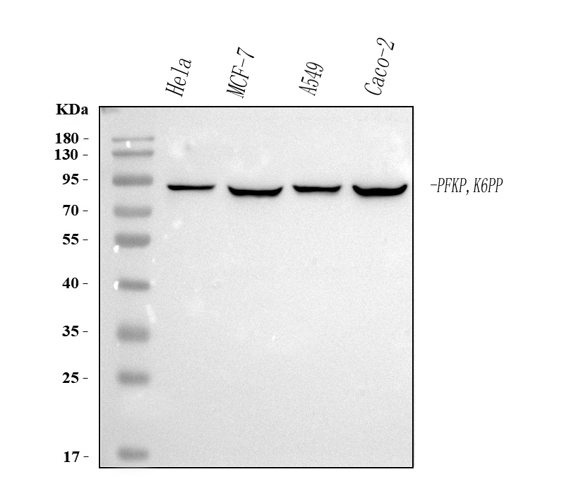
Western blot analysis of anti-PFKP antibody. The sample well of each lane was loaded with 30 ug of sample under reducing conditions. After electrophoresis, proteins were transferred to a membrane. Then the membrane was incubated with mouse anti-PFKP antigen affinity purified monoclonal antibody and probed with a goat anti-mouse IgG-HRP secondary antibody. The signal is developed using ECL Plus Western Blotting Substrate.IHC
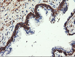
Immunohistochemical staining of paraffin-embedded Human prostate tissue within the normal limits using anti-PFKP mouse monoclonal antibody. (Heat-induced epitope retrieval by 10mM citric buffer, pH6.0, 100°C for 10min)ICC/IF
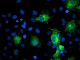
Anti-PFKP mouse monoclonal antibody immunofluorescent staining of COS7 cells transiently transfected by pCMV6-ENTRY PFKP .FC
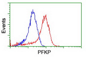
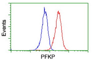
Flow cytometric Analysis of Hela cells, using anti-PFKP antibody, (Red), compared to a nonspecific negative control antibody, (Blue).FC
Flow cytometric Analysis of Jurkat cells, using anti-PFKP antibody, (Red), compared to a nonspecific negative control antibody, (Blue).| Product Name | Mouse monoclonal antibody to PFKP |
|---|---|
| Antibody Type | Primary Antibodies |
| Immunogen | Full length human recombinant protein of human PFKP(NP_002618) produced in HEK293T cell. |
| Clonality | Monoclonal |
|---|---|
| Isotype | IgG1 |
| Host Species | Mouse |
| Tested Applications | FCICC/IFIHCWB |
| WB:1:500-1:2000 IHC:1:150 ICC/IF:1:100 FC:1:100 |
|
| Species Reactivity | HumanMouseRat |
| Concentration | 0.5mg/ml |
| Purification | Protein A/G |
| Gene Symbol | PFKP |
|---|---|
| Gene Synonyms | PFKF PFK-C PFK-P ATP-PFK |
| Gene Full Name | phosphofructokinase, platelet |
| Gene Summary | This gene encodes a member of the phosphofructokinase A protein family. The encoded enzyme is the platelet-specific isoform of phosphofructokinase and plays a key role in glycolysis regulation. This gene may play a role in metabolic reprogramming in some cancers, including clear cell renal cell carcinomas, and cancer of the bladder, breast, and lung. Alternative splicing results in multiple transcript variants. [provided by RefSeq, Sep 2016] |
| Alternative Names | 6 phosphofructokinase type C PFK C PFKF PFKP Phosphofructokinase 1 phosphofructokinase platelet Phosphohexokinase |
| Molecular Weight(MW) | 86 kDa(Observed band size: 85.4 kDa) |
| Cellular Localization | Cytoplasm. |
WB
Western blot analysis of anti-PFKP antibody. The sample well of each lane was loaded with 30 ug of sample under reducing conditions. After electrophoresis, proteins were transferred to a membrane. Then the membrane was incubated with mouse anti-PFKP antigen affinity purified monoclonal antibody and probed with a goat anti-mouse IgG-HRP secondary antibody. The signal is developed using ECL Plus Western Blotting Substrate.
IHC
Immunohistochemical staining of paraffin-embedded Human prostate tissue within the normal limits using anti-PFKP mouse monoclonal antibody. (Heat-induced epitope retrieval by 10mM citric buffer, pH6.0, 100°C for 10min)
ICC/IF
Anti-PFKP mouse monoclonal antibody immunofluorescent staining of COS7 cells transiently transfected by pCMV6-ENTRY PFKP .
FC
Flow cytometric Analysis of Hela cells, using anti-PFKP antibody, (Red), compared to a nonspecific negative control antibody, (Blue).
FC
Flow cytometric Analysis of Jurkat cells, using anti-PFKP antibody, (Red), compared to a nonspecific negative control antibody, (Blue).| Application Notes | WB:1:500-1:2000 IHC:1:150 ICC/IF:1:100 FC:1:100 |
|---|
| Form | Liquid |
|---|---|
| Storage Instructions | Stable for 12 months from date of receipt. Store at -20°C as received. |
| Storage Buffer | PBS (PH 7.3) containing 1% BSA, 50% glycerol and 0.02% sodium azide. |


