WB
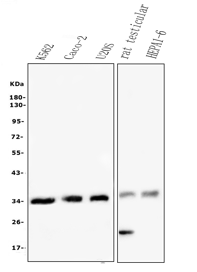
Western blot analysis of CD32a/FCGR2A using anti-CD32a/FCGR2A antibody. The sample well of each lane was loaded with 30 ug of sample under reducing conditions. After electrophoresis, proteins were transferred to a membrane. Then the membrane was incubated with rabbit anti-CD32a/FCGR2A antigen affinity purified polyclonal antibody at a dilution of 1:1000 and probed with a goat anti-rabbit IgG-HRP secondary antibody. The signal is developed using ECL Plus Western Blotting Substrate.IHC
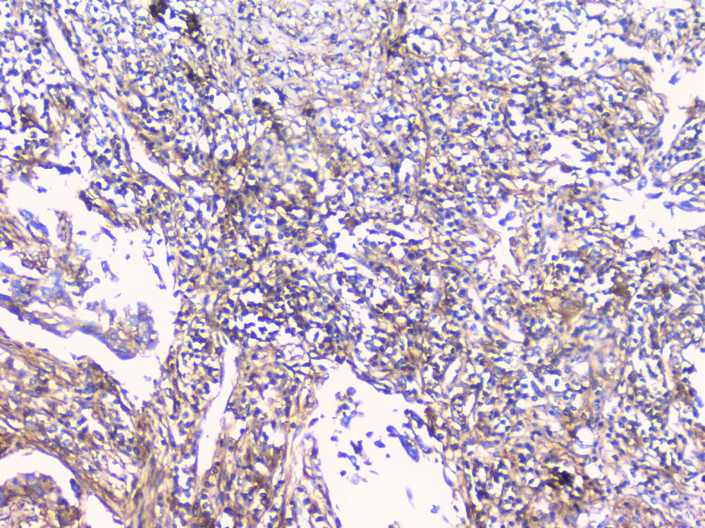
IHC analysis of CD32a/FCGR2A using anti-CD32a/FCGR2A antibody. CD32a/FCGR2A was detected in a paraffin-embedded section of human lung cancer tissue. Biotinylated goat anti-rabbit IgG was used as secondary antibody. The tissue section was incubated with rabbit anti-CD32a/FCGR2A Antibody at a dilution of 1:200 and developed using Strepavidin-Biotin-Complex (SABC) with DAB as the chromogen.IHC
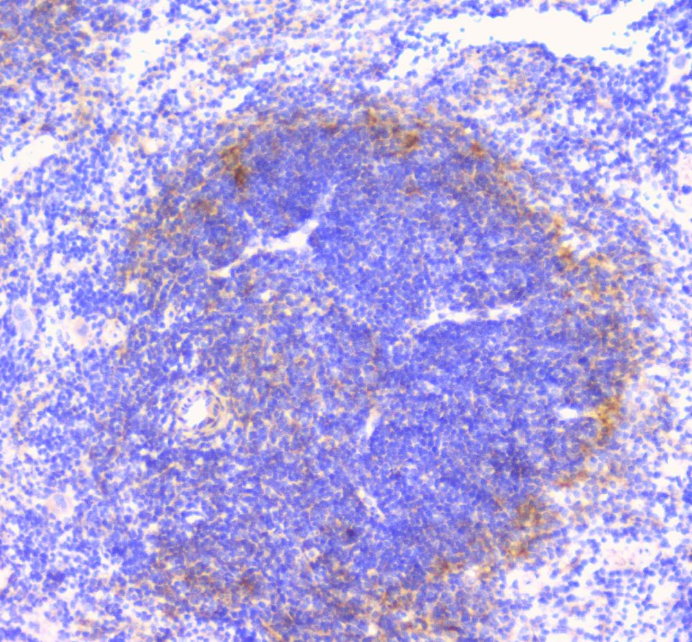
IHC analysis of CD32a/FCGR2A using anti-CD32a/FCGR2A antibody. CD32a/FCGR2A was detected in a paraffin-embedded section of mouse spleen tissue. Biotinylated goat anti-rabbit IgG was used as secondary antibody. The tissue section was incubated with rabbit anti-CD32a/FCGR2A Antibody at a dilution of 1:200 and developed using Strepavidin-Biotin-Complex (SABC) with DAB as the chromogen.IHC
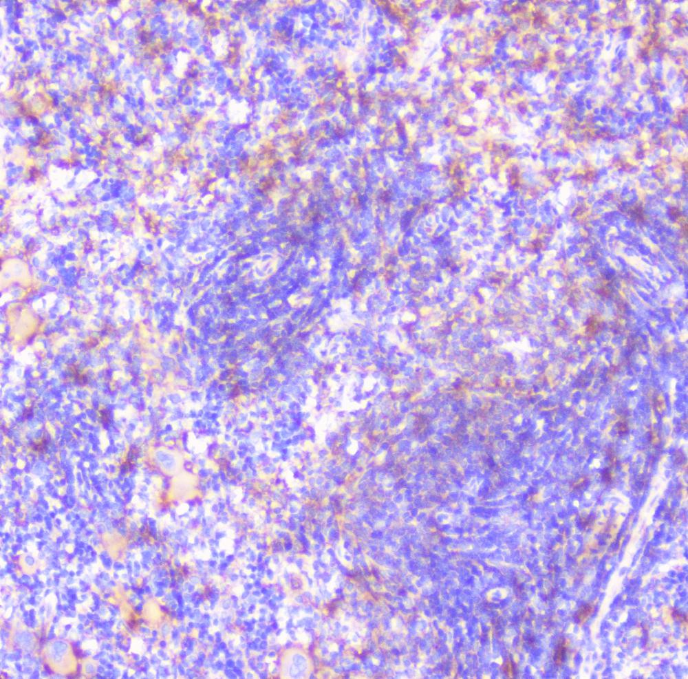
IHC analysis of CD32a/FCGR2A using anti-CD32a/FCGR2A antibody. CD32a/FCGR2A was detected in a paraffin-embedded section of rat spleen tissue. Biotinylated goat anti-rabbit IgG was used as secondary antibody. The tissue section was incubated with rabbit anti-CD32a/FCGR2A Antibody at a dilution of 1:200 and developed using Strepavidin-Biotin-Complex (SABC) with DAB as the chromogen.FC
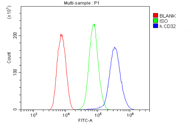
Flow Cytometry analysis of THP-1 cells using anti-FCGR2A antibody. Overlay histogram showing THP-1 cells stained with Rabbit polyclonal antibody to CD32a/FCGR2A (Blue line). anti-FCGR2A Antibody (1:100) for 30 min at 20°C. 488 conjugated goat anti-rabbit IgG (1:100) was used as secondary antibody . Isotype control antibody (Green line) was rabbit IgG (1:100) used under the same conditions. Unlabelled sample (Red line) was also used as a control.IF-P
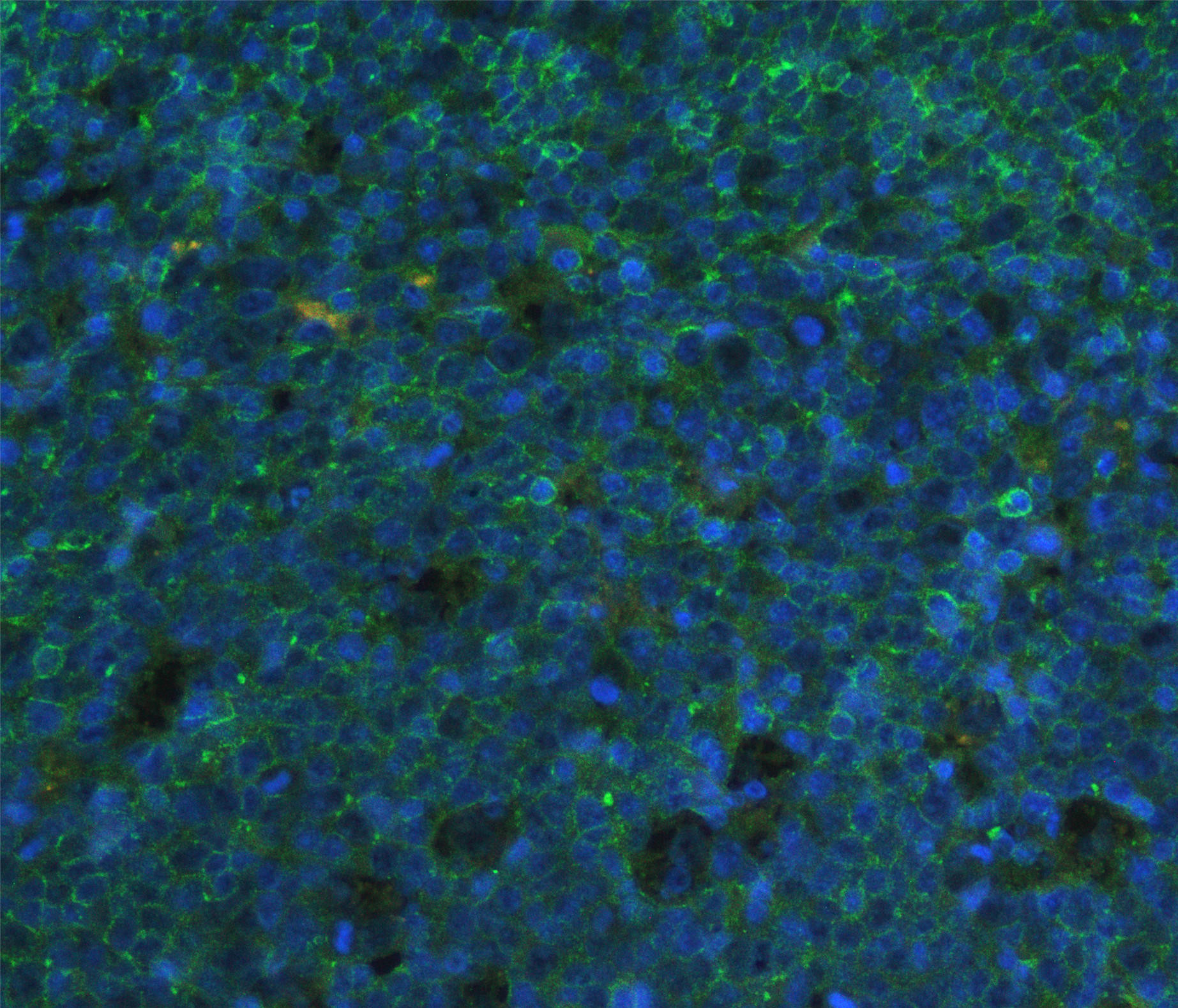
IF analysis of CD32 using anti-CD32 antibody. CD32 was detected in paraffin-embedded section of human tonsil tissues. Heat mediated antigen retrieval was performed in citrate buffer (pH6, epitope retrieval solution) for 20 mins. The tissue section was blocked with 10% goat serum. The tissue section was then incubated with 1μg/mL rabbit anti-CD32 Antibody overnight at 4°C. Fluoro488 Conjugated Goat Anti-Rabbit IgG was used as secondary antibody at 1:100 dilution and incubated for 30 minutes at 37°C. The section was counterstained with DAPI. Visualize using a fluorescence microscope and filter sets appropriate for the label used.| Product Name | Rabbit polyclonal antibody to CD32a/FCGR2A |
|---|---|
| Antibody Type | Primary Antibodies |
| Immunogen | E. coli-derived human FCGR2A recombinant protein (Position: Q34-M210). Human FCGR2A shares 64.1% and 60.7% amino acid (aa) sequence identity with mouse and rat FCGR2A, respectively. |
| Clonality | Polyclonal |
|---|---|
| Isotype | IgG |
| Host Species | Rabbit |
| Tested Applications | FCIF-PIHCWB |
| WB:1:500-1:2000 IHC:1:50-1:400 IF-P:1:50-400 FC:1:50-1:200 |
|
| Species Reactivity | HumanMouseRat |
| Concentration | 0.5mg/ml |
| Purification | Affinity purified |
| Gene Symbol | FCGR2A |
|---|---|
| Gene Synonyms | CD32 FCG2 FcGR CD32A CDw32 FCGR2 IGFR2 FCGR2A1 FcgammaRIIa |
| Gene Full Name | Fc gamma receptor IIa |
| Gene Summary | This gene encodes one member of a family of immunoglobulin Fc receptor genes found on the surface of many immune response cells. The protein encoded by this gene is a cell surface receptor found on phagocytic cells such as macrophages and neutrophils, and is involved in the process of phagocytosis and clearing of immune complexes. Alternative splicing results in multiple transcript variants. [provided by RefSeq, Oct 2008] |
| Alternative Names | CD32 CD32A CDw32 Fc gamma RII a Fc gamma RIIa FCG2 FcGR FCGR2 FCGR2A FCGR2A1 FcRII a IGFR2 IgG Fc receptor II a |
| Molecular Weight(MW) | 35 kDa |
| Cellular Localization | Cell membrane,Membrane. |
WB
Western blot analysis of CD32a/FCGR2A using anti-CD32a/FCGR2A antibody. The sample well of each lane was loaded with 30 ug of sample under reducing conditions. After electrophoresis, proteins were transferred to a membrane. Then the membrane was incubated with rabbit anti-CD32a/FCGR2A antigen affinity purified polyclonal antibody at a dilution of 1:1000 and probed with a goat anti-rabbit IgG-HRP secondary antibody. The signal is developed using ECL Plus Western Blotting Substrate.
IHC
IHC analysis of CD32a/FCGR2A using anti-CD32a/FCGR2A antibody. CD32a/FCGR2A was detected in a paraffin-embedded section of human lung cancer tissue. Biotinylated goat anti-rabbit IgG was used as secondary antibody. The tissue section was incubated with rabbit anti-CD32a/FCGR2A Antibody at a dilution of 1:200 and developed using Strepavidin-Biotin-Complex (SABC) with DAB as the chromogen.
IHC
IHC analysis of CD32a/FCGR2A using anti-CD32a/FCGR2A antibody. CD32a/FCGR2A was detected in a paraffin-embedded section of mouse spleen tissue. Biotinylated goat anti-rabbit IgG was used as secondary antibody. The tissue section was incubated with rabbit anti-CD32a/FCGR2A Antibody at a dilution of 1:200 and developed using Strepavidin-Biotin-Complex (SABC) with DAB as the chromogen.
IHC
IHC analysis of CD32a/FCGR2A using anti-CD32a/FCGR2A antibody. CD32a/FCGR2A was detected in a paraffin-embedded section of rat spleen tissue. Biotinylated goat anti-rabbit IgG was used as secondary antibody. The tissue section was incubated with rabbit anti-CD32a/FCGR2A Antibody at a dilution of 1:200 and developed using Strepavidin-Biotin-Complex (SABC) with DAB as the chromogen.
FC
Flow Cytometry analysis of THP-1 cells using anti-FCGR2A antibody. Overlay histogram showing THP-1 cells stained with Rabbit polyclonal antibody to CD32a/FCGR2A (Blue line). anti-FCGR2A Antibody (1:100) for 30 min at 20°C. 488 conjugated goat anti-rabbit IgG (1:100) was used as secondary antibody . Isotype control antibody (Green line) was rabbit IgG (1:100) used under the same conditions. Unlabelled sample (Red line) was also used as a control.
IF-P
IF analysis of CD32 using anti-CD32 antibody. CD32 was detected in paraffin-embedded section of human tonsil tissues. Heat mediated antigen retrieval was performed in citrate buffer (pH6, epitope retrieval solution) for 20 mins. The tissue section was blocked with 10% goat serum. The tissue section was then incubated with 1μg/mL rabbit anti-CD32 Antibody overnight at 4°C. Fluoro488 Conjugated Goat Anti-Rabbit IgG was used as secondary antibody at 1:100 dilution and incubated for 30 minutes at 37°C. The section was counterstained with DAPI. Visualize using a fluorescence microscope and filter sets appropriate for the label used.| Application Notes | WB:1:500-1:2000 IHC:1:50-1:400 IF-P:1:50-400 FC:1:50-1:200 |
|---|
| Form | Liquid |
|---|---|
| Storage Instructions | 12 months from date of receipt,-20℃ as supplied. |
| Storage Buffer | 500 ug/ml antibody with PBS, 0.02% NaN3, 1 mg/ml BSA and 50% glycerol. |


