WB
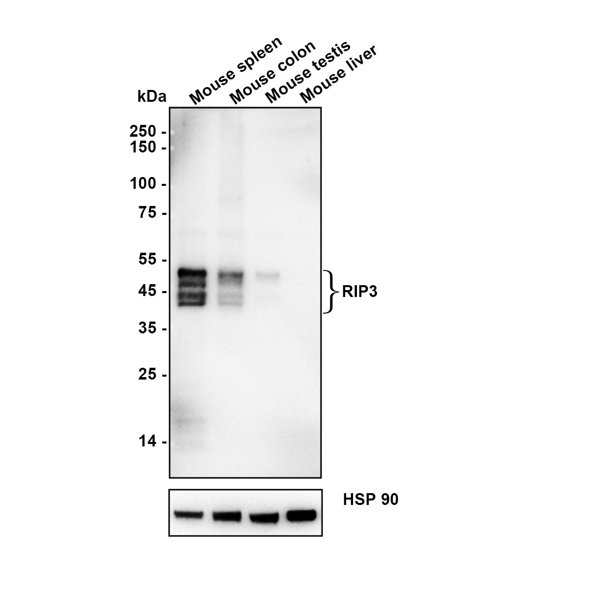
Relative expression (RE). Western blot analysis of RIP3 on different lysates with Rabbit anti-RIP3 antibody at 1/5,000 dilution. Lysates/proteins at 20 µg/Lane. Exposure time: 59 seconds; Low expression: liver ; Blocking: 5% NFDM/TBST, 1 hour at room temperature. Primary antibody: 1/5,000 in primary antibody dilution buffer, overnight at 4 ℃. Secondary antibody: Goat anti-Rabbit IgG-HRP, 1/50,000 in 5% NFDM/TBST, 1 hour at room temperature.IHC
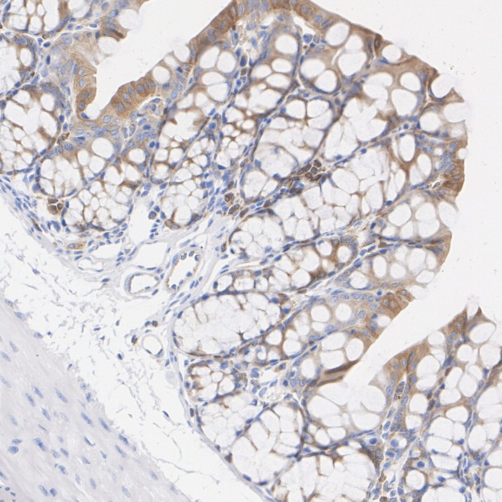
Application: Immunohistochemistry (IHC-P). Species: Mouse. Tissue: Colon. Sample: Paraffin-embedded section. Antigen retrieval: Heat-mediated, Tris-EDTA buffer (pH 9.0), 20 minutes at 95°C. Wash buffer: 1× TBST Endogenous peroxidase blocking: 3% H₂O₂, 10 minutes at room temperature. Blocking: 1% BSA + 10% normal goat serum, 10 minutes at room temperature. Primary antibody: 1/200, 1 hour at room temperature. Secondary antibody: 20 minutes at room temperature.ICC/IF
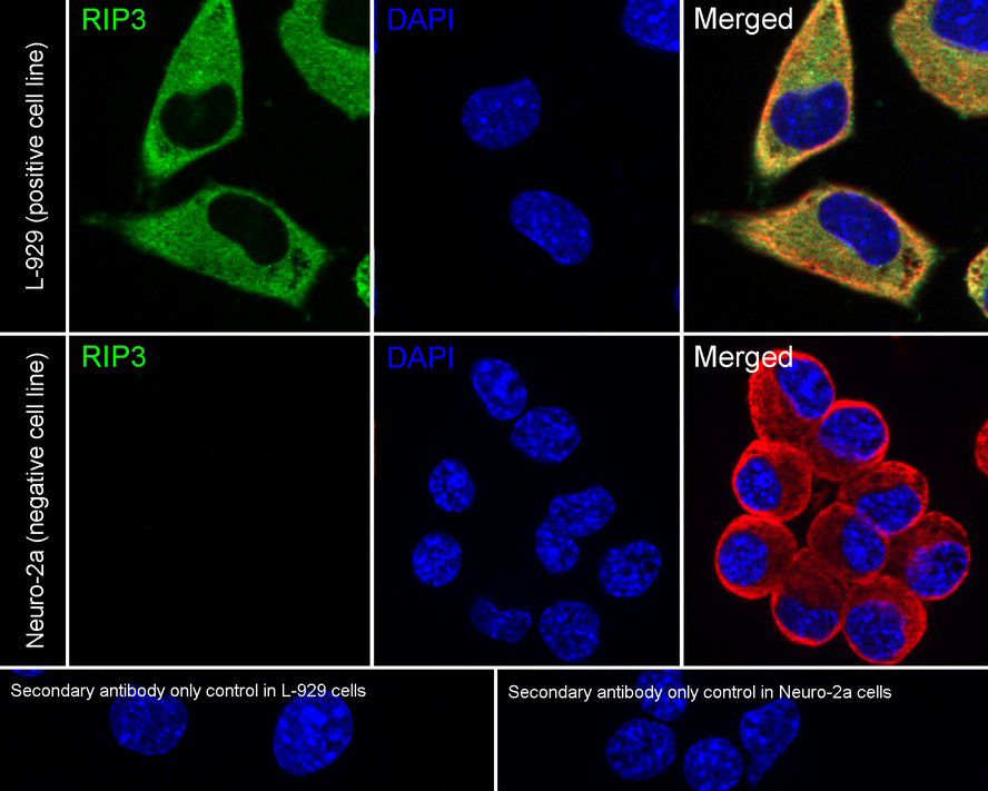
Relative expression (RE) Application: Immunocytochemistry (IF-cell) Species: Mouse Sample: L-929 (Mouse fibroblast)/Neuro-2a (Mouse brain neuroblastoma cell) Fixation: 4% Paraformaldehyde, 15 minutes at room temperature. Permeabilization: 0.1% Triton X-100, 15 minutes at room temperature. Blocking: 1% BSA + 10% normal goat serum, 1 hour at room temperature. Antibody dilution buffer: 1% BSA in PBST. Primary antibody: 1/100, overnight at 4°C. Secondary antibody: Goat Anti-Rabbit IgG (488), 45 minutes at room temperature. Counterstain: Beta tubulin (Red), 1/100, overnight at 4℃. The nuclear counterstain was DAPI (Blue). Negative control: Neuro-2a.FC
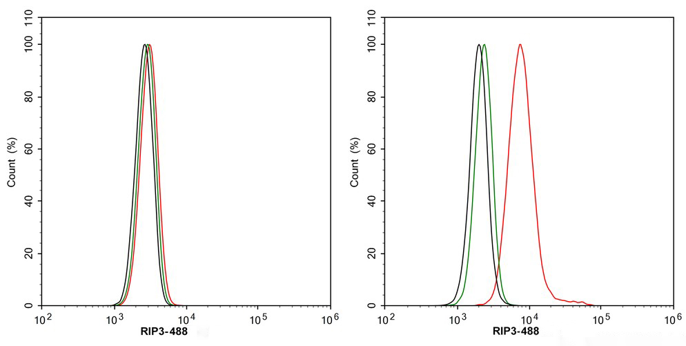
Relative expression (RE). Application: Flow Cytometry (Intra). Species: Mouse. Sample: Neuro-2a (Mouse brain neuroblastoma cells)/L-929 (Mouse fibroblast). Fixation: 4% Paraformaldehyde, 15 minutes at room temperature. Permeabilization: 0.1% Tween-20, 15 minutes at room temperature. Blocking: 1% BSA + 10% normal goat serum, 15 minutes at room temperature. Antibody dilution buffer: 1x PBS. Primary antibody: RIP3 Recombinant Rabbit Monoclonal Antibody (1/1,000, Red) compared with Rabbit IgG Isotype Control (Green), 15 minutes at room temperature. Secondary antibody: Goat Anti-Rabbit IgG (488), 15 minutes at room temperature. Negative control: Neuro-2a.IP
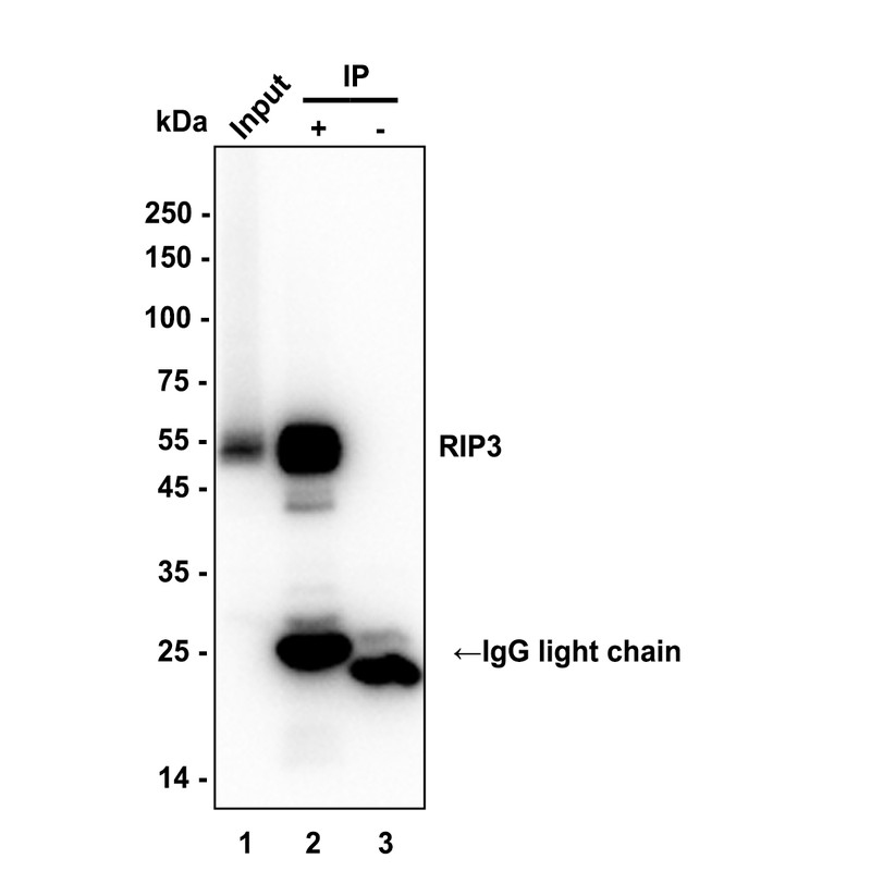
Immunoprecipitation (IP). RIP3 was immunoprecipitated in 0.2 mg L-929 (Mouse fibroblast) cell lysate cell lysate with RIP3 Recombinant Rabbit Monoclonal Antibody at 2 µg/10 µl beads. Western blot was performed from the immunoprecipitate using RIP3 Recombinant Rabbit Monoclonal Antibody at 1/5,000 dilution. HRP Conjugated Rabbit IgG kappa light chain Mouse Monoclonal Antibody at 1/5000 dilution was used for 1 hour at room temperature. Lane 1: L-929 cell lysate (input). Lane 2: RIP3 Recombinant Rabbit Monoclonal Antibody IP in L-929 cell lysate. Lane 3: Rabbit IgG instead ofRIP3 Recombinant Rabbit Monoclonal Antibody in L-929 cell lysate. Exposure time: 7 seconds Blocking: 5% NFDM/TBST, 1 hour at room temperature Primary dilution: 1/5,000 in primary antibody dilution buffer, 2 hours at room temperature.| Product Name | RIP3 Recombinant Rabbit Monoclonal Antibody |
|---|---|
| Antibody Type | Primary Antibodies |
| Immunogen | Recombinant protein within mouse RIP3 aa 301-486. |
| Clonality | Monoclonal |
|---|---|
| Isotype | IgG |
| Host Species | Rabbit |
| Tested Applications | FCICC/IFIHCIPWB |
| WB:1:5000 IHC:1:200 ICC/IF:1:100 FC:1:1000 IP:1-2μg/sample |
|
| Species Reactivity | Mouse |
| Concentration | 1mg/ml |
| Purification | Protein A |
| Gene Symbol | RIPK3 |
|---|---|
| Gene Synonyms | RIP3 |
| Gene Full Name | receptor interacting serine/threonine kinase 3 |
| Gene Summary | The product of this gene is a member of the receptor-interacting protein (RIP) family of serine/threonine protein kinases, and contains a C-terminal domain unique from other RIP family members. The encoded protein is predominantly localized to the cytoplasm, and can undergo nucleocytoplasmic shuttling dependent on novel nuclear localization and export signals. It is a component of the tumor necrosis factor (TNF) receptor-I signaling complex, and can induce apoptosis and weakly activate the NF-kappaB transcription factor. [provided by RefSeq, Jul 2008] |
| Alternative Names | Receptor interacting protein 3 antibody Receptor interacting serine threonine kinase 3 antibody Receptor interacting serine/threonine protein kinase 3 antibody Receptor-interacting protein 3 antibody Receptor-interacting serine/threonine-protein kinase 3 antibody RIP 3 antibody RIP like protein kinase 3 antibody RIP-3 antibody RIP-like protein kinase 3 antibody RIPK 3 antibody RIPK3 antibody |
| Molecular Weight(MW) | 53 kDa(Observed band size: 40-55 kDa) |
| Cellular Localization | Cytoplasm, cytosol, Nucleus. |
WB
Relative expression (RE). Western blot analysis of RIP3 on different lysates with Rabbit anti-RIP3 antibody at 1/5,000 dilution. Lysates/proteins at 20 µg/Lane. Exposure time: 59 seconds; Low expression: liver ; Blocking: 5% NFDM/TBST, 1 hour at room temperature. Primary antibody: 1/5,000 in primary antibody dilution buffer, overnight at 4 ℃. Secondary antibody: Goat anti-Rabbit IgG-HRP, 1/50,000 in 5% NFDM/TBST, 1 hour at room temperature.
IHC
Application: Immunohistochemistry (IHC-P). Species: Mouse. Tissue: Colon. Sample: Paraffin-embedded section. Antigen retrieval: Heat-mediated, Tris-EDTA buffer (pH 9.0), 20 minutes at 95°C. Wash buffer: 1× TBST Endogenous peroxidase blocking: 3% H₂O₂, 10 minutes at room temperature. Blocking: 1% BSA + 10% normal goat serum, 10 minutes at room temperature. Primary antibody: 1/200, 1 hour at room temperature. Secondary antibody: 20 minutes at room temperature.
ICC/IF
Relative expression (RE) Application: Immunocytochemistry (IF-cell) Species: Mouse Sample: L-929 (Mouse fibroblast)/Neuro-2a (Mouse brain neuroblastoma cell) Fixation: 4% Paraformaldehyde, 15 minutes at room temperature. Permeabilization: 0.1% Triton X-100, 15 minutes at room temperature. Blocking: 1% BSA + 10% normal goat serum, 1 hour at room temperature. Antibody dilution buffer: 1% BSA in PBST. Primary antibody: 1/100, overnight at 4°C. Secondary antibody: Goat Anti-Rabbit IgG (488), 45 minutes at room temperature. Counterstain: Beta tubulin (Red), 1/100, overnight at 4℃. The nuclear counterstain was DAPI (Blue). Negative control: Neuro-2a.
FC
Relative expression (RE). Application: Flow Cytometry (Intra). Species: Mouse. Sample: Neuro-2a (Mouse brain neuroblastoma cells)/L-929 (Mouse fibroblast). Fixation: 4% Paraformaldehyde, 15 minutes at room temperature. Permeabilization: 0.1% Tween-20, 15 minutes at room temperature. Blocking: 1% BSA + 10% normal goat serum, 15 minutes at room temperature. Antibody dilution buffer: 1x PBS. Primary antibody: RIP3 Recombinant Rabbit Monoclonal Antibody (1/1,000, Red) compared with Rabbit IgG Isotype Control (Green), 15 minutes at room temperature. Secondary antibody: Goat Anti-Rabbit IgG (488), 15 minutes at room temperature. Negative control: Neuro-2a.
IP
Immunoprecipitation (IP). RIP3 was immunoprecipitated in 0.2 mg L-929 (Mouse fibroblast) cell lysate cell lysate with RIP3 Recombinant Rabbit Monoclonal Antibody at 2 µg/10 µl beads. Western blot was performed from the immunoprecipitate using RIP3 Recombinant Rabbit Monoclonal Antibody at 1/5,000 dilution. HRP Conjugated Rabbit IgG kappa light chain Mouse Monoclonal Antibody at 1/5000 dilution was used for 1 hour at room temperature. Lane 1: L-929 cell lysate (input). Lane 2: RIP3 Recombinant Rabbit Monoclonal Antibody IP in L-929 cell lysate. Lane 3: Rabbit IgG instead ofRIP3 Recombinant Rabbit Monoclonal Antibody in L-929 cell lysate. Exposure time: 7 seconds Blocking: 5% NFDM/TBST, 1 hour at room temperature Primary dilution: 1/5,000 in primary antibody dilution buffer, 2 hours at room temperature.| Application Notes | WB:1:5000 IHC:1:200 ICC/IF:1:100 FC:1:1000 IP:1-2μg/sample |
|---|
| Form | Liquid |
|---|---|
| Storage Instructions | Shipped at 4℃. Store at +4℃ short term (1-2 weeks). Store at -20℃ long term. |
| Storage Buffer | 1*PBS (pH7.4), 0.1% BSA, 40% Glycerol, 0.2% Proclean 950. |


