WB
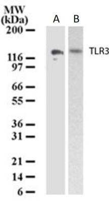
Western Blot: TLR3 Antibody - Analysis of TLR3 in lysates from A) human intestine and F) ovary using TLR3 antibody at 3 ug/ml. Goat anti-mouse HRP conjugate was used as secondary.IHC
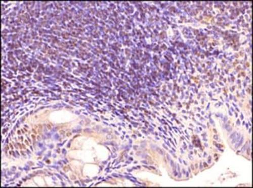
Immunohistochemistry-Paraffin: TLR3 Antibody - Tissue section of mouse intestine using TLR3 antibody at 1:500 dilution with HRP-DAB detection and hematoxylin counterstaining. The representative image shows a punctate staining of the ER and endosomes in a subset of cells in Peyer's patches (organized lymphoid nodules) in the tested section.IHC
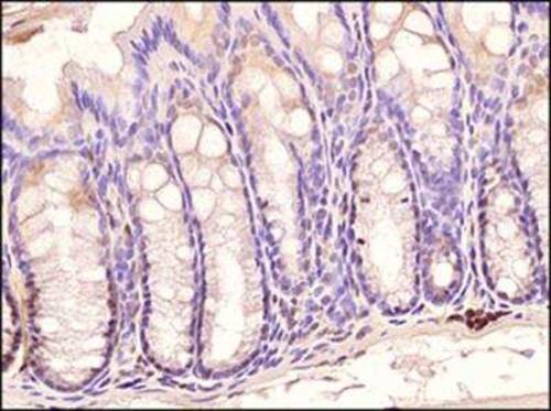
Immunohistochemistry-Paraffin: TLR3 Antibody - Mouse colon using TLR3 antibody at 1:500 dilution with HRP-DAB detection and hematoxylin counterstaining. Intense signal was found in subset of cells at the bases of the crypts.| Product Name | TLR3 Mouse Monoclonal Antibody |
|---|---|
| Antibody Type | Primary Antibodies |
| Immunogen | A synthetic peptide corresponding to amino acids 55-85 of human TLR3 was used as immunogen. |
| Clonality | Monoclonal |
|---|---|
| Isotype | IgG1, k |
| Host Species | Mouse |
| Tested Applications | IHCWB |
| WB:1-3 ug/ml IHC:1:10-1:500 |
|
| Species Reactivity | CanineHumanMouse |
| Concentration | 1mg/ml |
| Purification | Protein G |
| Gene Symbol | TLR3 |
|---|---|
| Gene Synonyms | CD283 IIAE2 IMD83 |
| Gene Full Name | toll like receptor 3 |
| Gene Summary | The protein encoded by this gene is a member of the Toll-like receptor (TLR) family which plays a fundamental role in pathogen recognition and activation of innate immunity. TLRs are highly conserved from Drosophila to humans and share structural and functional similarities. They recognize pathogen-associated molecular patterns (PAMPs) that are expressed on infectious agents, and mediate the production of cytokines necessary for the development of effective immunity. The various TLRs exhibit different patterns of expression. This receptor is most abundantly expressed in placenta and pancreas, and is restricted to the dendritic subpopulation of the leukocytes. It recognizes dsRNA associated with viral infection, and induces the activation of NF-kappaB and the production of type I interferons. It thus plays a role in host defense against multiple viruses. [provided by RefSeq, Jul 2021] |
| Alternative Names | CD283 IIAE2 |
| Cellular Localization | Endoplasmic reticulum,Endosome,Membrane. |
WB
Western Blot: TLR3 Antibody - Analysis of TLR3 in lysates from A) human intestine and F) ovary using TLR3 antibody at 3 ug/ml. Goat anti-mouse HRP conjugate was used as secondary.
IHC
Immunohistochemistry-Paraffin: TLR3 Antibody - Tissue section of mouse intestine using TLR3 antibody at 1:500 dilution with HRP-DAB detection and hematoxylin counterstaining. The representative image shows a punctate staining of the ER and endosomes in a subset of cells in Peyer's patches (organized lymphoid nodules) in the tested section.
IHC
Immunohistochemistry-Paraffin: TLR3 Antibody - Mouse colon using TLR3 antibody at 1:500 dilution with HRP-DAB detection and hematoxylin counterstaining. Intense signal was found in subset of cells at the bases of the crypts.| Application Notes | WB:1-3 ug/ml IHC:1:10-1:500 |
|---|
| Form | Liquid |
|---|---|
| Storage Instructions | Store at -20°C as received. Store at 4°C short term. Aliquot and store at -20°C long term. Avoid freeze-thaw cycles. |
| Storage Buffer | PBS containing 0.05% BSA, 0.05% Sodium Azide. |


