IHC
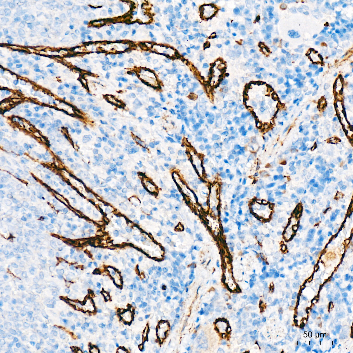
Immunohistochemistry analysis of paraffin embedded Rat spleen tissue using ZO-1 Rabbit mAb at a dilution of 1:1000 (40x lens). High pressure antigen retrieval was performed with 0.01 M citrate buffer (pH 6.0) prior to IHC stainingIHC
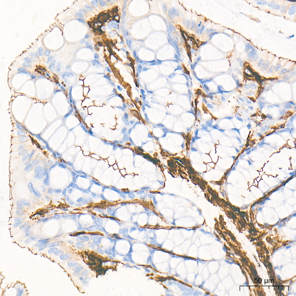
Immunohistochemistry analysis of paraffin embedded Mouse intestine tissue using ZO-1 Rabbit mAb at a dilution of 1:800 (40x lens). High pressure antigen retrieval performed with 0.01M Tris-EDTA Buffer (pH 9.0) prior to IHC staining.IF-P
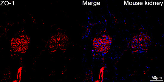
Confocal imaging of paraffin-embedded Mouse kidney tissue using ZO-1 Rabbit mAb (dilution 1:200) followed by a further incubation with Cy3 Goat Anti-Rabbit IgG (H+L) (dilution 1:500) (Red). DAPI was used for nuclear staining (Blue). High pressure antigen retrieval performed with 0.01M Citrate Buffer(pH 6.0) prior to IF staining. Objective: 40x.IF-P
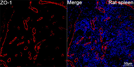
Confocal imaging of paraffin-embedded Rat spleen tissue using ZO-1 Rabbit mAb (dilution 1:200) followed by a further incubation with Cy3 Goat Anti-Rabbit IgG (H+L) (dilution 1:500) (Red). DAPI was used for nuclear staining (Blue). High pressure antigen retrieval performed with 0.01M Citrate Buffer(pH 6.0) prior to IF staining. Objective: 40x.ICC/IF
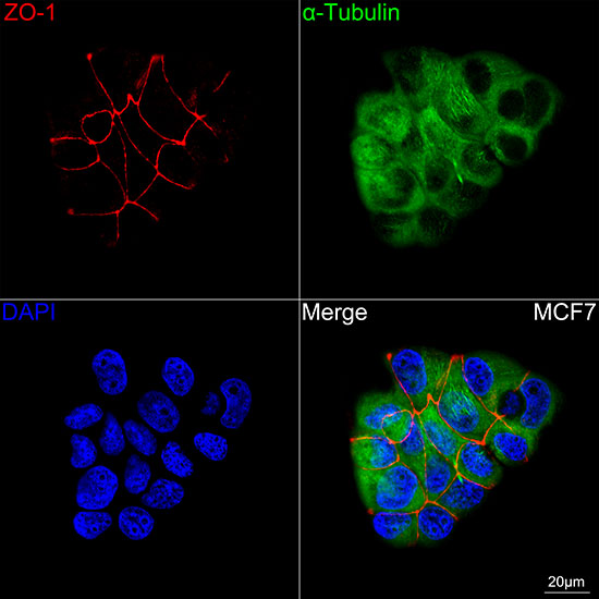
Confocal imaging of MCF7 cells using ZO-1 Rabbit mAb (dilution 1:100) followed by a further incubation with Cy3 Goat Anti-Rabbit IgG (H+L) (dilution 1:500) (Red). The cells were counterstained with α-Tubulin Mouse mAb (dilution 1:400) followed by incubation with 488-conjugated Goat Anti-Mouse IgG (H+L) Ab (dilution 1:500) (Green). DAPI was used for nuclear staining (Blue). Objective: 100x.| Product Name | ZO-1 Rabbit mAb |
|---|---|
| Antibody Type | Primary Antibodies |
| Immunogen | Recombinant protein. |
| Clonality | Monoclonal |
|---|---|
| Isotype | IgG |
| Host Species | Rabbit |
| Tested Applications | ICC/IFIF-PIHC |
| IHC:1:500-1:1000 IF-P:1:100-1:800 ICC/IF:1:100-1:800 |
|
| Species Reactivity | HumanMouseRat |
| Concentration | 1mg/ml |
| Purification | Affinity purified |
| Gene Symbol | TJP1 |
|---|---|
| Gene Synonyms | ZO-1 |
| Gene Full Name | tight junction protein 1 |
| Gene Summary | This gene encodes a member of the membrane-associated guanylate kinase (MAGUK) family of proteins, and acts as a tight junction adaptor protein that also regulates adherens junctions. Tight junctions regulate the movement of ions and macromolecules between endothelial and epithelial cells. The multidomain structure of this scaffold protein, including a postsynaptic density 95/disc-large/zona occludens (PDZ) domain, a Src homology (SH3) domain, a guanylate kinase (GuK) domain and unique (U) motifs all help to co-ordinate binding of transmembrane proteins, cytosolic proteins, and F-actin, which are required for tight junction function. Alternative splicing results in multiple transcript variants encoding different isoforms. [provided by RefSeq, Aug 2017] |
| Molecular Weight(MW) | 195 kDa |
| Cellular Localization | Cell junction, Cell membrane, Cytoplasmic side, Peripheral membrane protein, gap junction, tight junction. |
IHC
Immunohistochemistry analysis of paraffin embedded Rat spleen tissue using ZO-1 Rabbit mAb at a dilution of 1:1000 (40x lens). High pressure antigen retrieval was performed with 0.01 M citrate buffer (pH 6.0) prior to IHC staining
IHC
Immunohistochemistry analysis of paraffin embedded Mouse intestine tissue using ZO-1 Rabbit mAb at a dilution of 1:800 (40x lens). High pressure antigen retrieval performed with 0.01M Tris-EDTA Buffer (pH 9.0) prior to IHC staining.
IF-P
Confocal imaging of paraffin-embedded Mouse kidney tissue using ZO-1 Rabbit mAb (dilution 1:200) followed by a further incubation with Cy3 Goat Anti-Rabbit IgG (H+L) (dilution 1:500) (Red). DAPI was used for nuclear staining (Blue). High pressure antigen retrieval performed with 0.01M Citrate Buffer(pH 6.0) prior to IF staining. Objective: 40x.
IF-P
Confocal imaging of paraffin-embedded Rat spleen tissue using ZO-1 Rabbit mAb (dilution 1:200) followed by a further incubation with Cy3 Goat Anti-Rabbit IgG (H+L) (dilution 1:500) (Red). DAPI was used for nuclear staining (Blue). High pressure antigen retrieval performed with 0.01M Citrate Buffer(pH 6.0) prior to IF staining. Objective: 40x.
ICC/IF
Confocal imaging of MCF7 cells using ZO-1 Rabbit mAb (dilution 1:100) followed by a further incubation with Cy3 Goat Anti-Rabbit IgG (H+L) (dilution 1:500) (Red). The cells were counterstained with α-Tubulin Mouse mAb (dilution 1:400) followed by incubation with 488-conjugated Goat Anti-Mouse IgG (H+L) Ab (dilution 1:500) (Green). DAPI was used for nuclear staining (Blue). Objective: 100x.| Application Notes | IHC:1:500-1:1000 IF-P:1:100-1:800 ICC/IF:1:100-1:800 |
|---|
| Form | Liquid |
|---|---|
| Storage Instructions | Store at -20℃. Avoid freeze / thaw cycles. |
| Storage Buffer | PBS with 0.01% thimerosal, 50% glycerol, pH7.3. |


