WB
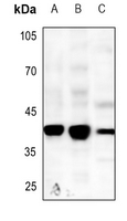
Western blot analysis of Caspase 5 expression in HCT116 (A), CT26 (B), C6 (C) whole cell lysates.IHC
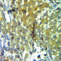
Immunohistochemical analysis of Caspase 5 staining in human prostate formalin fixed paraffin embedded tissue section. The section was pre-treated using heat mediated antigen retrieval with sodium citrate buffer (pH 6.0). The section was then incubated with the antibody at room temperature and detected using an HRP conjugated compact polymer system. DAB was used as the chromogen. The section was then counterstained with haematoxylin and mounted with DPX.ICC/IF
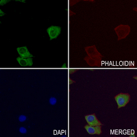
Immunofluorescent analysis of Caspase 5 staining in HepG2 cells. Formalin-fixed cells were permeabilized with 0.1% Triton X-100 in TBS for 5-10 minutes and blocked with 3% BSA-PBS for 30 minutes at room temperature. Cells were probed with the primary antibody in 3% BSA-PBS and incubated overnight at 4 °C in a hidified chamber. Cells were washed with PBST and incubated with a AF488-conjugated secondary antibody (green) in PBS at room temperature in the dark. Phalloidin - AF594 was used to stain Actin filaments (red). DAPI was used to stain the cell nuclei (blue).| Product Name | Anti-Caspase 5 Antibody |
|---|---|
| Antibody Type | Primary Antibodies |
| Immunogen | KLH-conjugated synthetic peptide encompassing a sequence within the center region of human Caspase 5. The exact sequence is proprietary. |
| Clonality | Polyclonal |
|---|---|
| Isotype | IgG |
| Host Species | Rabbit |
| Tested Applications | ICC/IFIHCWB |
| WB:1:500-1:1000 IHC:1:50-1:100 ICC/IF:1:50-1:200 |
|
| Species Reactivity | HumanMouseRat |
| Concentration | 1mg/ml |
| Purification | Affinity purified |
| Gene Symbol | CASP5 |
|---|---|
| Gene Synonyms | ICH-3 ICEREL-III ICE(rel)III |
| Gene Full Name | caspase 5 |
| Gene Summary | This gene encodes a member of the cysteine-aspartic acid protease (caspase) family. Sequential activation of caspases plays a central role in the execution-phase of cell apoptosis. Caspases exist as inactive proenzymes which undergo proteolytic processing at conserved aspartic residues to produce two subunits, large and small, that dimerize to form the active enzyme. Overexpression of the active form of this enzyme induces apoptosis in fibroblasts. Max, a central component of the Myc/Max/Mad transcription regulation network important for cell growth, differentiation, and apoptosis, is cleaved by this protein; this process requires Fas-mediated dephosphorylation of Max. The expression of this gene is regulated by interferon-gamma and lipopolysaccharide. Alternatively spliced transcript variants have been identified for this gene. [provided by RefSeq, Aug 2010] |
| Alternative Names | ICH3 Caspase-5 CASP-5 ICE(rel)-III Protease ICH-3 Protease TY |
| Molecular Weight(MW) | 49 kDa(Observed band size: 35;44;50 kDa) |
WB
Western blot analysis of Caspase 5 expression in HCT116 (A), CT26 (B), C6 (C) whole cell lysates.
IHC
Immunohistochemical analysis of Caspase 5 staining in human prostate formalin fixed paraffin embedded tissue section. The section was pre-treated using heat mediated antigen retrieval with sodium citrate buffer (pH 6.0). The section was then incubated with the antibody at room temperature and detected using an HRP conjugated compact polymer system. DAB was used as the chromogen. The section was then counterstained with haematoxylin and mounted with DPX.
ICC/IF
Immunofluorescent analysis of Caspase 5 staining in HepG2 cells. Formalin-fixed cells were permeabilized with 0.1% Triton X-100 in TBS for 5-10 minutes and blocked with 3% BSA-PBS for 30 minutes at room temperature. Cells were probed with the primary antibody in 3% BSA-PBS and incubated overnight at 4 °C in a hidified chamber. Cells were washed with PBST and incubated with a AF488-conjugated secondary antibody (green) in PBS at room temperature in the dark. Phalloidin - AF594 was used to stain Actin filaments (red). DAPI was used to stain the cell nuclei (blue).| Application Notes | WB:1:500-1:1000 IHC:1:50-1:100 ICC/IF:1:50-1:200 |
|---|
| Form | Liquid |
|---|---|
| Storage Instructions | Shipped at 4°C. Upon delivery aliquot and store at -20°C for one year. Avoid freeze/thaw cycles. |
| Storage Buffer | Liquid in 0.42% Potassium phosphate, 0.87% Sodium chloride, pH 7.3, 30% glycerol, and 0.01% sodium azide. |


