WB
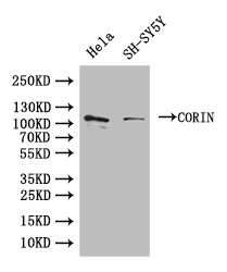
Western Blot. Positive WB detected in: Hela whole cell lysate,SH-SY5Y whole cell lysate. All lanes: CORIN antibody at 1:1000. Secondary Goat polyclonal to rabbit IgG at 1/50000 dilution.IHC
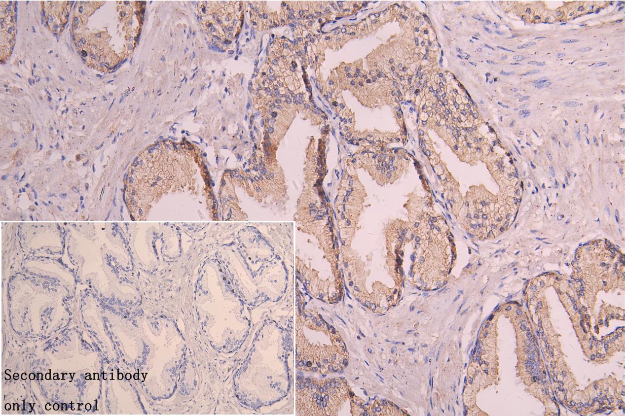
IHC image of Rabbit anti-CORIN Polyclonal antibody diluted at 1:150 and staining in paraffin-embedded Human prostate cancer performed on a Leica BondTM system. After dewaxing and hydration, antigen retrieval was mediated by high pressure in a citrate buffer (pH 6.0). Section was blocked with 10% normal goat serum 30min at RT. Then primary antibody (1% BSA) was incubated at 4°C overnight. The primary is detected by a Goat anti-rabbit polymer IgG labeled by HRP and visualized using 0.05% DAB.ICC/IF
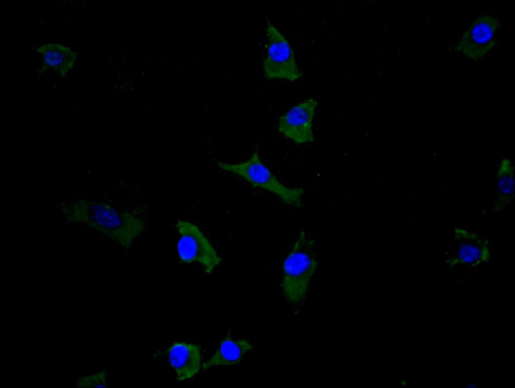
Immunofluorescence staining of SH-SY5Y cell with Rabbit anti-CORIN Polyclonal antibody at 1:30, counter-stained with DAPI. The cells were fixed in 4% formaldehyde and blocked in 10% normal Goat Serum. The cells were then incubated with the antibody overnight at 4°C. The secondary antibody was Alexa Fluor 488-congugated AffiniPure Goat Anti-Rabbit IgG(H+L).| Product Name | Rabbit anti-CORIN Polyclonal antibody |
|---|---|
| Antibody Type | Primary Antibodies |
| Immunogen | Recombinant Human Atrial natriuretic peptide-converting enzyme protein. |
| Clonality | Polyclonal |
|---|---|
| Isotype | IgG |
| Host Species | Rabbit |
| Tested Applications | ICC/IFIHCWB |
| WB:1:1000-1:3000 IHC:1:100-1:300 ICC/IF:1:20-1:200 |
|
| Species Reactivity | Human |
| Concentration | 1mg/ml |
| Purification | Affinity purified |
| Gene Symbol | CORIN |
|---|---|
| Gene Synonyms | CRN ATC2 Lrp4 PEE5 CMH30 TMPRSS10 |
| Gene Full Name | corin, serine peptidase |
| Gene Summary | This gene encodes a member of the type II transmembrane serine protease class of the trypsin superfamily. Members of this family are composed of multiple structurally distinct domains. The encoded protein converts pro-atrial natriuretic peptide to biologically active atrial natriuretic peptide, a cardiac hormone that regulates blood volume and pressure. This protein may also function as a pro-brain-type natriuretic peptide convertase. Multiple alternatively spliced transcript variants encoding different isoforms have been found for this gene. [provided by RefSeq, Jun 2013] |
| Alternative Names | activated protease fragment antibody ATC 2 antibody ATC2 antibody Atrial natriuretic peptide-converting enzyme antibody Atrial natriuteric peptide converting enzyme antibody Corin antibody Corin serine peptidase antibody CORIN_HUMAN antibody CRN antibody Heart specific serine proteinase antibody Heart specific serine proteinase ATC2 antibody Heart-specific serine proteinase ATC2 antibody MGC119742 antibody Pro ANP convertase antibody Pro ANP converting enzyme antibody Pro-ANP-converting enzyme antibody TMPRSS 10 antibody TMPRSS10 antibody Transmembrane protease serine 10 antibody |
| Molecular Weight(MW) | 117 kDa |
| Cellular Localization | Cell membrane,Membrane,Secreted. |
WB
Western Blot. Positive WB detected in: Hela whole cell lysate,SH-SY5Y whole cell lysate. All lanes: CORIN antibody at 1:1000. Secondary Goat polyclonal to rabbit IgG at 1/50000 dilution.
IHC
IHC image of Rabbit anti-CORIN Polyclonal antibody diluted at 1:150 and staining in paraffin-embedded Human prostate cancer performed on a Leica BondTM system. After dewaxing and hydration, antigen retrieval was mediated by high pressure in a citrate buffer (pH 6.0). Section was blocked with 10% normal goat serum 30min at RT. Then primary antibody (1% BSA) was incubated at 4°C overnight. The primary is detected by a Goat anti-rabbit polymer IgG labeled by HRP and visualized using 0.05% DAB.
ICC/IF
Immunofluorescence staining of SH-SY5Y cell with Rabbit anti-CORIN Polyclonal antibody at 1:30, counter-stained with DAPI. The cells were fixed in 4% formaldehyde and blocked in 10% normal Goat Serum. The cells were then incubated with the antibody overnight at 4°C. The secondary antibody was Alexa Fluor 488-congugated AffiniPure Goat Anti-Rabbit IgG(H+L).| Application Notes | WB:1:1000-1:3000 IHC:1:100-1:300 ICC/IF:1:20-1:200 |
|---|
| Form | Liquid |
|---|---|
| Storage Instructions | Upon receipt, store at -20°C or -80°C. Avoid repeated freeze. |
| Storage Buffer | PBS with 50% glycerol, pH7.4. |


