WB
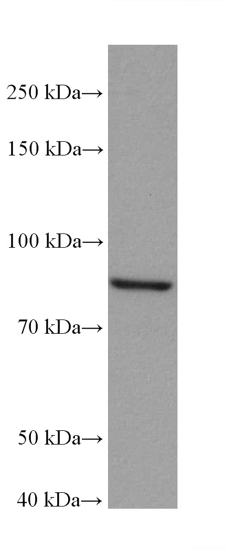
WB analysis of HEK-293 using Aconitase 2 Monoclonal antibody. HEK-293 cells were subjected to SDS PAGE followed by western blot with ACO2 antibody at dilution of 1:20000 incubated at room temperature for 1.5 hours.WB
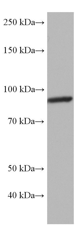
WB analysis of mouse brain using Aconitase 2 Monoclonal antibody. mouse brain tissue were subjected to SDS PAGE followed by western blot with ACO2 antibody at dilution of 1:3000 incubated at room temperature for 1.5 hours.WB
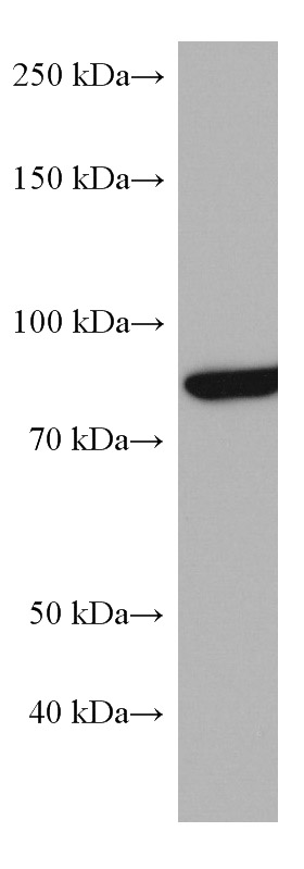
WB analysis of pig brain using Aconitase 2 Monoclonal antibody. pig brain tissue were subjected to SDS PAGE followed by western blot with ACO2 antibody at dilution of 1:3000 incubated at room temperature for 1.5 hours.WB
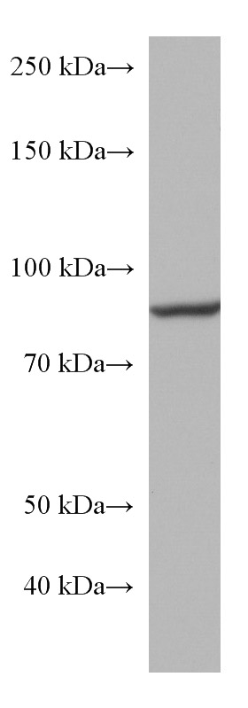
WB analysis of rat brain using Aconitase 2 Monoclonal antibody. rat brain tissue were subjected to SDS PAGE followed by western blot with ACO2 antibody at dilution of 1:3000 incubated at room temperature for 1.5 hours.IHC
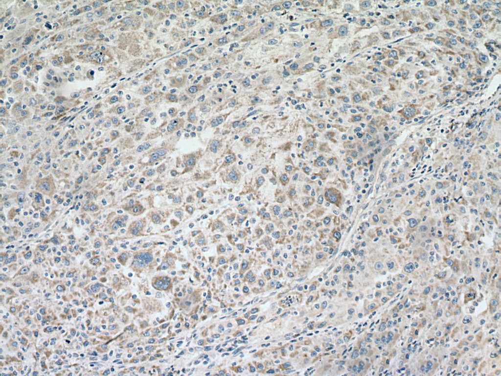
IHC staining of human liver cancer using Aconitase 2 Monoclonal antibody. Immunohistochemical analysis of paraffin-embedded human liver cancer tissue slide using ACO2 antibody at dilution of 1:2000 (under 10x lens). Heat mediated antigen retrieval with Tris-EDTA buffer (pH 9.0).FC
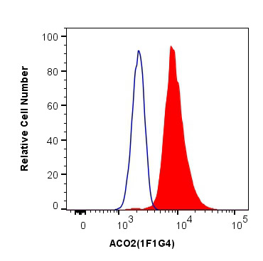
FC experiment of HeLa using Aconitase 2 Monoclonal antibody. 1X10^6 HeLa cells were intracellularly stained with 0.4 ug Anti-Human ACO2 and 488-Conjugated AffiniPure Goat Anti-Mouse IgG(H+L) at dilution 1:1000 (red), or 0.4 ug Mouse IgG1 Isotype Control (blue). Cells were fixed with 4% PFA and permeabilized with Flow Cytometry Perm Buffer.| Product Name | Aconitase 2 Monoclonal antibody |
|---|---|
| Antibody Type | Primary Antibodies |
| Immunogen | Recombinant human ACO2 protein. |
| Clonality | Monoclonal |
|---|---|
| Isotype | IgG1 |
| Host Species | Mouse |
| Tested Applications | FCIHCWB |
| WB:1:5000-1:50000 IHC:1:500-1:2000 FC:0.40 ug per 10^6 cells in a 100 µl suspension |
|
| Species Reactivity | HumanMousePigRat |
| Concentration | 1mg/ml |
| Purification | Protein G |
| Gene Symbol | ACO2 |
|---|---|
| Gene Synonyms | ICRD OCA8 OPA9 ACONM HEL-S-284 |
| Gene Full Name | aconitase 2 |
| Gene Summary | The protein encoded by this gene belongs to the aconitase/IPM isomerase family. It is an enzyme that catalyzes the interconversion of citrate to isocitrate via cis-aconitate in the second step of the TCA cycle. This protein is encoded in the nucleus and functions in the mitochondrion. It was found to be one of the mitochondrial matrix proteins that are preferentially degraded by the serine protease 15(PRSS15), also known as Lon protease, after oxidative modification. [provided by RefSeq, Jul 2008] |
| Alternative Names | ACO2 1F1G4 ACO 2 Aconitase Aconitase2 |
| Molecular Weight(MW) | 85 kDa |
| Cellular Localization | Mitochondrion. |
WB
WB analysis of HEK-293 using Aconitase 2 Monoclonal antibody. HEK-293 cells were subjected to SDS PAGE followed by western blot with ACO2 antibody at dilution of 1:20000 incubated at room temperature for 1.5 hours.
WB
WB analysis of mouse brain using Aconitase 2 Monoclonal antibody. mouse brain tissue were subjected to SDS PAGE followed by western blot with ACO2 antibody at dilution of 1:3000 incubated at room temperature for 1.5 hours.
WB
WB analysis of pig brain using Aconitase 2 Monoclonal antibody. pig brain tissue were subjected to SDS PAGE followed by western blot with ACO2 antibody at dilution of 1:3000 incubated at room temperature for 1.5 hours.
WB
WB analysis of rat brain using Aconitase 2 Monoclonal antibody. rat brain tissue were subjected to SDS PAGE followed by western blot with ACO2 antibody at dilution of 1:3000 incubated at room temperature for 1.5 hours.
IHC
IHC staining of human liver cancer using Aconitase 2 Monoclonal antibody. Immunohistochemical analysis of paraffin-embedded human liver cancer tissue slide using ACO2 antibody at dilution of 1:2000 (under 10x lens). Heat mediated antigen retrieval with Tris-EDTA buffer (pH 9.0).
FC
FC experiment of HeLa using Aconitase 2 Monoclonal antibody. 1X10^6 HeLa cells were intracellularly stained with 0.4 ug Anti-Human ACO2 and 488-Conjugated AffiniPure Goat Anti-Mouse IgG(H+L) at dilution 1:1000 (red), or 0.4 ug Mouse IgG1 Isotype Control (blue). Cells were fixed with 4% PFA and permeabilized with Flow Cytometry Perm Buffer.| Application Notes | WB:1:5000-1:50000 IHC:1:500-1:2000 FC:0.40 ug per 10^6 cells in a 100 µl suspension |
|---|
| Form | Liquid |
|---|---|
| Storage Instructions | Store at -20°C. Stable for one year after shipment. Aliquoting is unnecessary for -20°C storage. |
| Storage Buffer | PBS with 0.02% sodium azide and 50% glycerol, pH 7.3. |


