WB
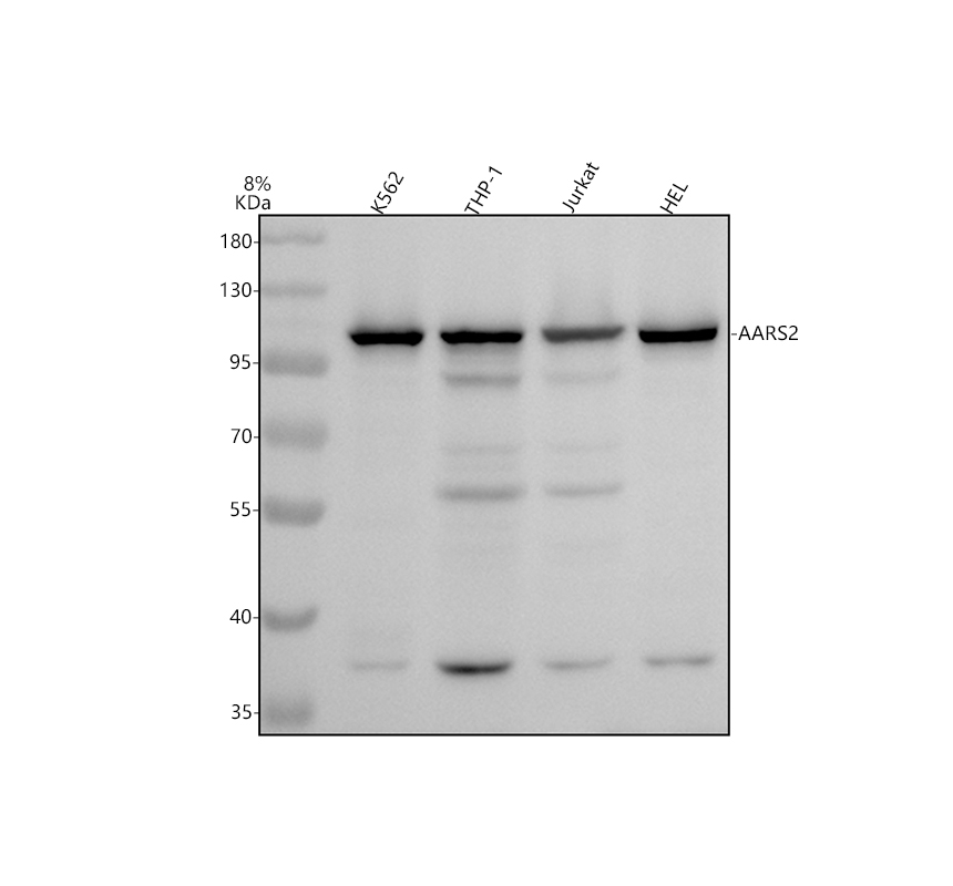
Western blot analysis of AARS2 using anti-AARS2 antibody. The sample well of each lane was loaded with 30 ug of sample under reducing conditions. After electrophoresis, proteins were transferred to a membrane. Then the membrane was incubated with rabbit anti-AARS2 antigen affinity purified polyclonal antibody at a dilution of 1:1000 and probed with a goat anti-rabbit IgG-HRP secondary antibody. The signal is developed using ECL Plus Western Blotting Substrate.IHC
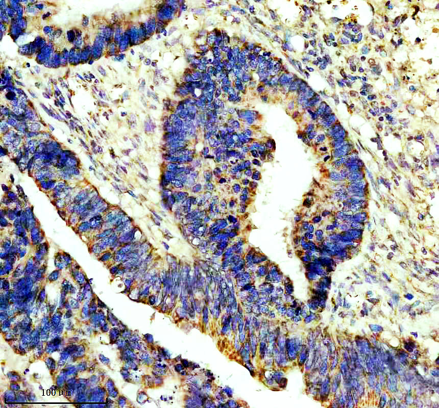
IHC analysis of AARS2 using anti-AARS2 antibody. AARS2 was detected in a paraffin-embedded section of human colon cancer tissue. The tissue section was incubated with rabbit anti-AARS2 Antibody at a dilution of 1:200 and developed using HRP Conjugated Rabbit IgG Super Vision Assay Kit with DAB as the chromogen.IHC
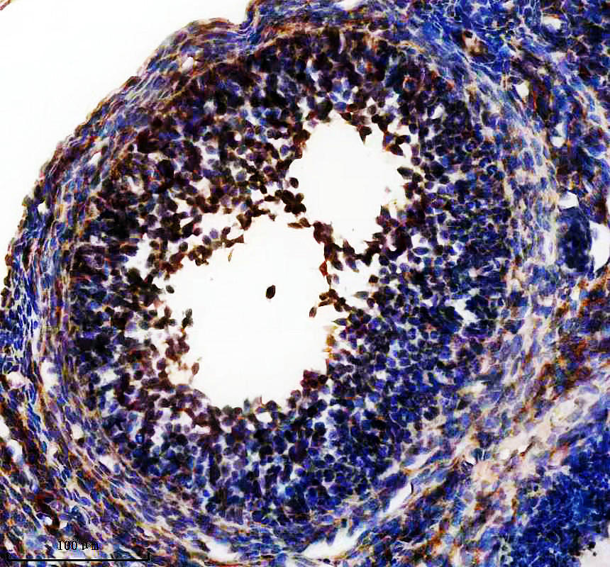
IHC analysis of AARS2 using anti-AARS2 antibody. AARS2 was detected in a paraffin-embedded section of rat ovary tissue. The tissue section was incubated with rabbit anti-AARS2 Antibody at a dilution of 1:200 and developed using HRP Conjugated Rabbit IgG Super Vision Assay Kit with DAB as the chromogen.IHC
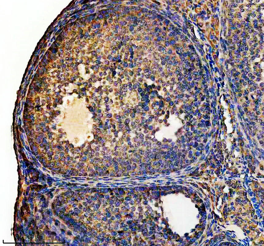
IHC analysis of AARS2 using anti-AARS2 antibody. AARS2 was detected in a paraffin-embedded section of mouse ovary tissue. The tissue section was incubated with rabbit anti-AARS2 Antibody at a dilution of 1:200 and developed using HRP Conjugated Rabbit IgG Super Vision Assay Kit with DAB as the chromogen.| Product Name | Rabbit polyclonal antibody to AARS2 |
|---|---|
| Antibody Type | Primary Antibodies |
| Immunogen | E.coli-derived human AARS2 recombinant protein (Position: L637-L982). |
| Clonality | Polyclonal |
|---|---|
| Isotype | IgG |
| Host Species | Rabbit |
| Tested Applications | IHCWB |
| WB:1:1000-1:5000 IHC:1:50-1:400 |
|
| Species Reactivity | HumanMouseRat |
| Concentration | 0.5mg/ml |
| Purification | Affinity purified |
| Gene Symbol | AARS2 |
|---|---|
| Gene Synonyms | AARSL LKENP COXPD8 MTALARS MT-ALARS |
| Gene Full Name | alanyl-tRNA synthetase 2, mitochondrial |
| Gene Summary | The protein encoded by this gene belongs to the class-II aminoacyl-tRNA synthetase family. Aminoacyl-tRNA synthetases play critical roles in mRNA translation by charging tRNAs with their cognate amino acids. The encoded protein is a mitochondrial enzyme that specifically aminoacylates alanyl-tRNA. Mutations in this gene are a cause of combined oxidative phosphorylation deficiency 8. [provided by RefSeq, Dec 2011] |
| Alternative Names | AARS2 AARSL Alanine tRNA ligase Alanyl tRNA synthetase like AlaRS KIAA1270 MT ALARS MTALARS |
| Molecular Weight(MW) | 107 kDa(Observed band size: 105-115 kDa) |
| Cellular Localization | Mitochondrion. |
WB
Western blot analysis of AARS2 using anti-AARS2 antibody. The sample well of each lane was loaded with 30 ug of sample under reducing conditions. After electrophoresis, proteins were transferred to a membrane. Then the membrane was incubated with rabbit anti-AARS2 antigen affinity purified polyclonal antibody at a dilution of 1:1000 and probed with a goat anti-rabbit IgG-HRP secondary antibody. The signal is developed using ECL Plus Western Blotting Substrate.
IHC
IHC analysis of AARS2 using anti-AARS2 antibody. AARS2 was detected in a paraffin-embedded section of human colon cancer tissue. The tissue section was incubated with rabbit anti-AARS2 Antibody at a dilution of 1:200 and developed using HRP Conjugated Rabbit IgG Super Vision Assay Kit with DAB as the chromogen.
IHC
IHC analysis of AARS2 using anti-AARS2 antibody. AARS2 was detected in a paraffin-embedded section of rat ovary tissue. The tissue section was incubated with rabbit anti-AARS2 Antibody at a dilution of 1:200 and developed using HRP Conjugated Rabbit IgG Super Vision Assay Kit with DAB as the chromogen.
IHC
IHC analysis of AARS2 using anti-AARS2 antibody. AARS2 was detected in a paraffin-embedded section of mouse ovary tissue. The tissue section was incubated with rabbit anti-AARS2 Antibody at a dilution of 1:200 and developed using HRP Conjugated Rabbit IgG Super Vision Assay Kit with DAB as the chromogen.| Application Notes | WB:1:1000-1:5000 IHC:1:50-1:400 |
|---|
| Form | Liquid |
|---|---|
| Storage Instructions | 12 months from date of receipt,-20℃ as supplied. |
| Storage Buffer | 500 ug/ml antibody with PBS, 0.02% NaN3, 1 mg/ml BSA and 50% glycerol. |


