WB
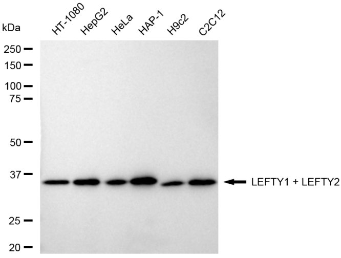
Western blotting analysis using LEFTY1 + LEFTY2 antibody. Total cell lysates (30 μg) from various cell lines were loaded and separated by SDS-PAGE. The blot was incubated with LEFTY1 + LEFTY2 antibody and HRP-conjugated goat anti-rabbit secondary antibody respectively.ICC/IF
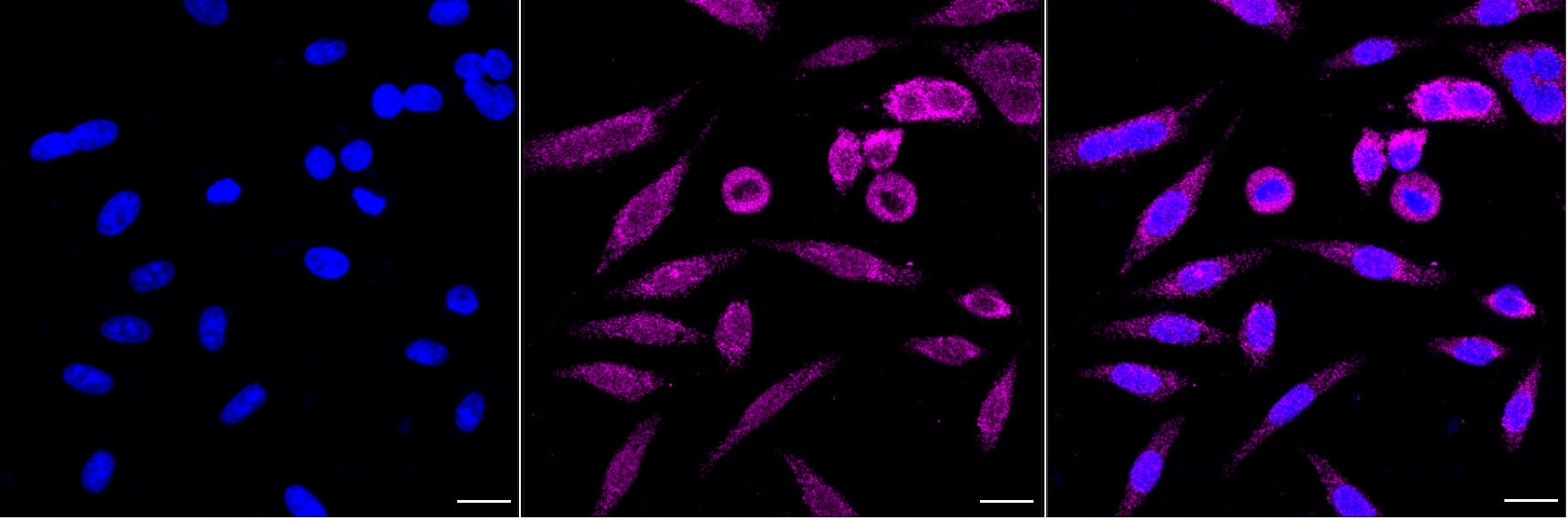
Immunocytochemical staining of HepG2 cells with LEFTY1 + LEFTY2 antibody. Nuclei were stained blue with DAPI; LEFTY1 + LEFTY2 was stained magenta with Alexa 647. Images were taken using Leica stellaris 5. Protein abundance based on laser Intensity and smart gain: Low. Scale bar, 20 μm.| Product Name | LEFTY1/LEFTY2 Recombinant Monoclonal Antibody |
|---|---|
| Antibody Type | Primary Antibodies |
| Immunogen | A synthesized peptide derived from Human LEFTY1/LEFTY2 protein. |
| Clonality | Monoclonal |
|---|---|
| Isotype | IgG |
| Host Species | Rabbit |
| Tested Applications | ICC/IFWB |
| WB:1:1000-1:5000 ICC/IF:1:100-1:1000 |
|
| Species Reactivity | HumanMouseRat |
| Concentration | 1mg/ml |
| Purification | Affinity purified |
| Gene Symbol | LEFTY2 |
|---|---|
| Gene Synonyms | EBAF LEFTA TGFB4 LEFTYA |
| Gene Full Name | left-right determination factor 2 |
| Gene Summary | This gene encodes a secreted ligand of the TGF-beta (transforming growth factor-beta) superfamily of proteins. Ligands of this family bind various TGF-beta receptors leading to recruitment and activation of SMAD family transcription factors that regulate gene expression. The encoded preproprotein is proteolytically processed to generate the mature protein, which plays a role in left-right asymmetry determination of organ systems during development. The protein may also play a role in endometrial bleeding. Mutations in this gene have been associated with left-right axis malformations, particularly in the heart and lungs. Some types of infertility have been associated with dysregulated expression of this gene in the endometrium. This gene is closely linked to both a related family member and a related pseudogene. This gene encodes multiple isoforms that may undergo similar proteolytic processing. [provided by RefSeq, Aug 2016] |
| Alternative Names | LEFTY1 Left-Right Determination Factor 1 LEFTYB LEFTB Left-Right Determination Factor B Protein Lefty-1 Protein Lefty-B Left-Right Determination Factor B LEFTY2 Left-Right Determination Factor 2 LEFTYA LEFTA TGFB4 EBAF Endometrial Bleeding-Associated Factor Left-Right Determination Factor A Transforming Growth Factor Beta-4 Protein Lefty-2 Protein Lefty-A TGF-Beta-4 Endometrial Bleeding Associated Factor (Left-Right Determination Factor A Transforming Growth Factor Beta Superfamily) Transforming Growth Factor Beta-4 (Endometrial Bleeding-Associated Factor LEFTY A) |
| Cellular Localization | Secreted. |
WB
Western blotting analysis using LEFTY1 + LEFTY2 antibody. Total cell lysates (30 μg) from various cell lines were loaded and separated by SDS-PAGE. The blot was incubated with LEFTY1 + LEFTY2 antibody and HRP-conjugated goat anti-rabbit secondary antibody respectively.
ICC/IF
Immunocytochemical staining of HepG2 cells with LEFTY1 + LEFTY2 antibody. Nuclei were stained blue with DAPI; LEFTY1 + LEFTY2 was stained magenta with Alexa 647. Images were taken using Leica stellaris 5. Protein abundance based on laser Intensity and smart gain: Low. Scale bar, 20 μm.| Application Notes | WB:1:1000-1:5000 ICC/IF:1:100-1:1000 |
|---|
| Form | Liquid |
|---|---|
| Storage Instructions | Upon receipt, store at -20°C or -80°C. Avoid repeated freeze. |
| Storage Buffer | Rabbit IgG in PBS (pH 7.4) containing 50% glycerol, and 0.02% sodium azide. |


