WB
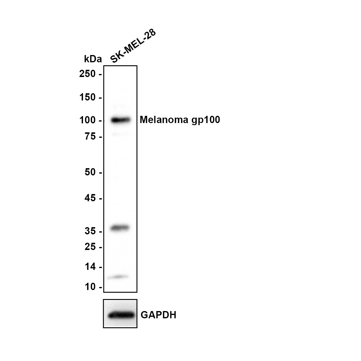
Western blot analysis of Melanoma gp100 on SK-MEL-28 cell lysates with Rabbit anti-Melanoma gp100 antibody at 1/1,000 dilution. Lysates/proteins at 20 µg/Lane. Exposure time: 2 minutes.IHC
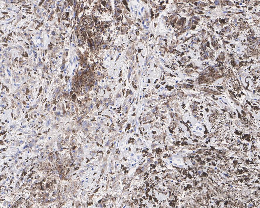
Immunohistochemical analysis of paraffin-embedded human melanoma tissue with Rabbit anti-Melanoma gp100 antibody at 1/1,000 dilution. The section was pre-treated using heat mediated antigen retrieval with Tris-EDTA buffer (pH 9.0) for 20 minutes. The tissues were blocked in 1% BSA for 20 minutes at room temperature, washed with ddH2O and PBS, and then probed with the primary antibody at 1/1,000 dilution for 1 hour at room temperature. The detection was performed using an HRP conjugated compact polymer system. DAB was used as the chromogen. Tissues were counterstained with hematoxylin and mounted with DPX.IHC
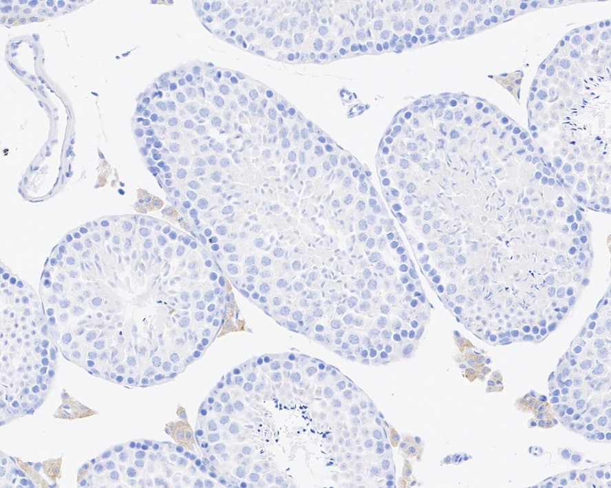
Immunohistochemical analysis of paraffin-embedded mouse testis tissue with Rabbit anti-Melanoma gp100 antibody at 1/200 dilution. The section was pre-treated using heat mediated antigen retrieval with Tris-EDTA buffer (pH 9.0) for 20 minutes. The tissues were blocked in 1% BSA for 20 minutes at room temperature, washed with ddH2O and PBS, and then probed with the primary antibody at 1/200 dilution for 1 hour at room temperature. The detection was performed using an HRP conjugated compact polymer system. DAB was used as the chromogen. Tissues were counterstained with hematoxylin and mounted with DPX.ICC/IF
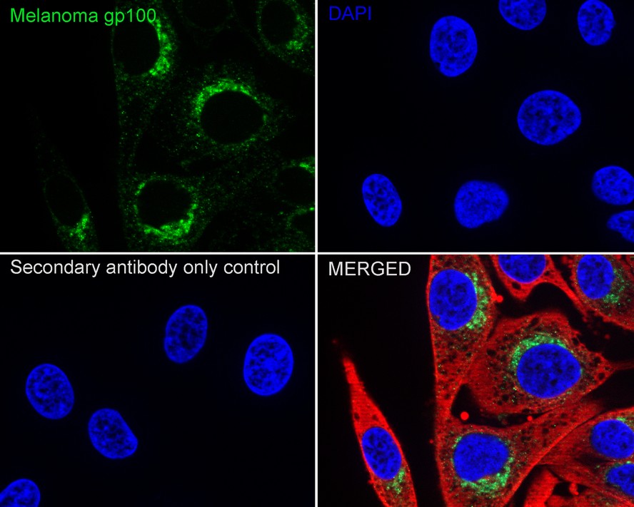
Immunocytochemistry analysis of SK-MEL-28 cells labeling Melanoma gp100 with Rabbit anti-Melanoma gp100 antibody at 1/100 dilution. Cells were fixed in 4% paraformaldehyde for 15 minutes at room temperature, permeabilized with 0.1% Triton X-100 in PBS for 15 minutes at room temperature, then blocked with 1% BSA in 10% negative goat serum for 1 hour at room temperature. Cells were then incubated with Rabbit anti-Melanoma gp100 antibody at 1/100 dilution in 1% BSA in PBST overnight at 4 ℃. Goat Anti-Rabbit IgG H&L (488) was used as the secondary antibody at 1/1,000 dilution. PBS instead of the primary antibody was used as the secondary antibody only control. Nuclear DNA was labelled in blue with DAPI. Beta tubulin (red) was stained at 1/100 dilution overnight at +4℃. Goat Anti-Mouse IgG H&L (594) was used as the secondary antibody at 1/1,000 dilution.| Product Name | Melanoma gp100 Recombinant Rabbit Monoclonal Antibody |
|---|---|
| Antibody Type | Primary Antibodies |
| Immunogen | Synthetic peptide within Human Melanoma gp100 aa 617-661 / 661. |
| Clonality | Monoclonal |
|---|---|
| Isotype | IgG |
| Host Species | Rabbit |
| Tested Applications | ICC/IFIHCWB |
| WB:1:1000 IHC:1:1000 ICC/IF:1:100 |
|
| Species Reactivity | HumanMouse |
| Concentration | 1mg/ml |
| Purification | Protein A |
| Gene Symbol | PMEL |
|---|---|
| Gene Synonyms | P1 SI SIL ME20 P100 SILV HMB45 ME20M gp100 HMB-45 ME20-M PMEL17 D12S53E |
| Gene Full Name | premelanosome protein |
| Gene Summary | This gene encodes a melanocyte-specific type I transmembrane glycoprotein. The encoded protein is enriched in melanosomes, which are the melanin-producing organelles in melanocytes, and plays an essential role in the structural organization of premelanosomes. This protein is involved in generating internal matrix fibers that define the transition from Stage I to Stage II melanosomes. This protein undergoes a complex pattern of prosttranslational processing and modification that is essential to the proper functioning of the protein. A secreted form of this protein that is released by proteolytic ectodomain shedding may be used as a melanoma-specific serum marker. Alternate splicing results in multiple transcript variants. [provided by RefSeq, Jan 2011] |
| Alternative Names | 95 kDa melanocyte specific secreted glycoprotein antibody 95 kDa melanocyte-specific secreted glycoprotein antibody D12S53E antibody gp100 antibody M-beta antibody ME20 antibody ME20 M/ME20 S antibody ME20-M antibody ME20-S antibody ME20M antibody ME20M/ME20S antibody ME20S antibody Melanocyte lineage specific antigen GP100 antibody Melanocyte protein mel 17 antibody Melanocyte protein Pmel 17 antibody Melanocyte protein Pmel 17 precursor antibody Melanocytes lineage-specific antigen GP100 antibody Melanoma associated ME20 antigen antibody Melanoma gp100 antibody Melanoma-associated ME20 antigen antibody Melanosomal matrix protein 17 antibody Melanosomal matrix protein17 antibody P1 antibody p100 antibody p26 antibody PMEL 17 antibody PMEL antibody PMEL_HUMAN antibody PMEL17 antibody Premelanosome protein antibody Secreted melanoma-associated ME20 antigen antibody SI antibody SIL antibody SILV antibody Silver (mouse homolog) like antibody Silver homolog antibody Silver locus protein homolog antibody Silver mouse homolog of antibody Melanosome(HMB-45) |
| Molecular Weight(MW) | 70 kDa(Observed band size: 100 kDa) |
| Cellular Localization | Endoplasmic reticulum membrane, Golgi apparatus, cis-Golgi network membrane, Endosome, multivesicular body, Melanosome, Extracellular vesicle, Secreted. |
WB
Western blot analysis of Melanoma gp100 on SK-MEL-28 cell lysates with Rabbit anti-Melanoma gp100 antibody at 1/1,000 dilution. Lysates/proteins at 20 µg/Lane. Exposure time: 2 minutes.
IHC
Immunohistochemical analysis of paraffin-embedded human melanoma tissue with Rabbit anti-Melanoma gp100 antibody at 1/1,000 dilution. The section was pre-treated using heat mediated antigen retrieval with Tris-EDTA buffer (pH 9.0) for 20 minutes. The tissues were blocked in 1% BSA for 20 minutes at room temperature, washed with ddH2O and PBS, and then probed with the primary antibody at 1/1,000 dilution for 1 hour at room temperature. The detection was performed using an HRP conjugated compact polymer system. DAB was used as the chromogen. Tissues were counterstained with hematoxylin and mounted with DPX.
IHC
Immunohistochemical analysis of paraffin-embedded mouse testis tissue with Rabbit anti-Melanoma gp100 antibody at 1/200 dilution. The section was pre-treated using heat mediated antigen retrieval with Tris-EDTA buffer (pH 9.0) for 20 minutes. The tissues were blocked in 1% BSA for 20 minutes at room temperature, washed with ddH2O and PBS, and then probed with the primary antibody at 1/200 dilution for 1 hour at room temperature. The detection was performed using an HRP conjugated compact polymer system. DAB was used as the chromogen. Tissues were counterstained with hematoxylin and mounted with DPX.
ICC/IF
Immunocytochemistry analysis of SK-MEL-28 cells labeling Melanoma gp100 with Rabbit anti-Melanoma gp100 antibody at 1/100 dilution. Cells were fixed in 4% paraformaldehyde for 15 minutes at room temperature, permeabilized with 0.1% Triton X-100 in PBS for 15 minutes at room temperature, then blocked with 1% BSA in 10% negative goat serum for 1 hour at room temperature. Cells were then incubated with Rabbit anti-Melanoma gp100 antibody at 1/100 dilution in 1% BSA in PBST overnight at 4 ℃. Goat Anti-Rabbit IgG H&L (488) was used as the secondary antibody at 1/1,000 dilution. PBS instead of the primary antibody was used as the secondary antibody only control. Nuclear DNA was labelled in blue with DAPI. Beta tubulin (red) was stained at 1/100 dilution overnight at +4℃. Goat Anti-Mouse IgG H&L (594) was used as the secondary antibody at 1/1,000 dilution.| Application Notes | WB:1:1000 IHC:1:1000 ICC/IF:1:100 |
|---|
| Form | Liquid |
|---|---|
| Storage Instructions | Shipped at 4℃. Store at +4℃ short term (1-2 weeks). Store at -20℃ long term. |
| Storage Buffer | 1*PBS (pH7.4), 0.1% BSA, 40% Glycerol, 0.2% Proclean 950. |


