WB
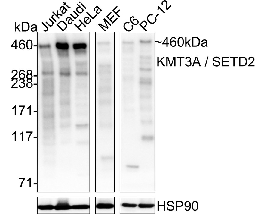
Western blot analysis of KMT3A / SETD2 on different lysates with Rabbit anti-KMT3A / SETD2 antibody at 1/5,000 dilution. Lysates/proteins at 20 µg/Lane. Exposure time: 30 seconds; 3-8% SDS-PAGE gel. Proteins were transferred to a PVDF membrane and blocked with 5% NFDM/TBST for 1 hour at room temperature. The primary antibody at 1/5,000 dilution was used in primary antibody dilution at 4℃ overnight. Goat Anti-Rabbit IgG - HRP Secondary Antibody at 1/50,000 dilution was used for 1 hour at room temperature.IHC
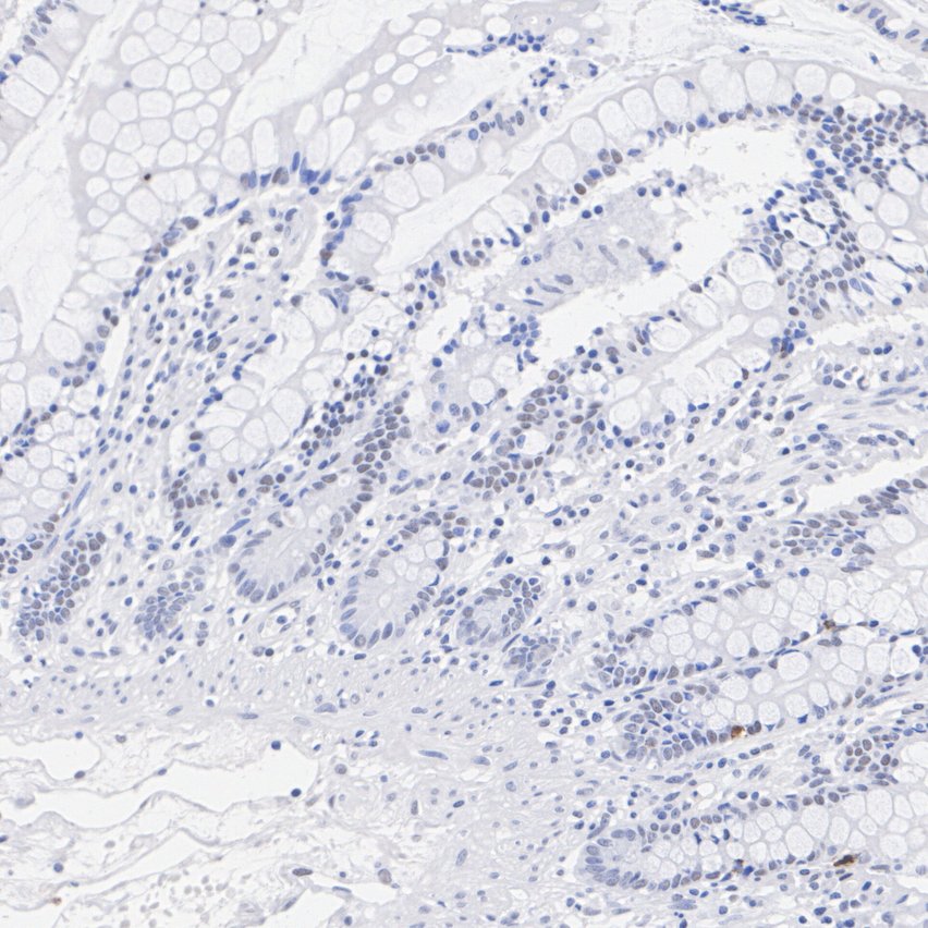
Immunohistochemical analysis of paraffin-embedded human small intestine tissue with Rabbit anti-KMT3A / SETD2 antibody at 1/200 dilution. The section was pre-treated using heat mediated antigen retrieval with Tris-EDTA buffer (pH 9.0) for 20 minutes. The tissues were blocked in 1% BSA for 20 minutes at room temperature, washed with ddH2O and PBS, and then probed with the primary antibody at 1/200 dilution for 1 hour at room temperature. The detection was performed using an HRP conjugated compact polymer system. DAB was used as the chromogen. Tissues were counterstained with hematoxylin and mounted with DPX.IHC
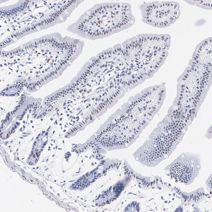
Immunohistochemical analysis of paraffin-embedded mouse small intestine tissue with Rabbit anti-KMT3A / SETD2 antibody at 1/200 dilution. The section was pre-treated using heat mediated antigen retrieval with Tris-EDTA buffer (pH 9.0) for 20 minutes. The tissues were blocked in 1% BSA for 20 minutes at room temperature, washed with ddH2O and PBS, and then probed with the primary antibody at 1/200 dilution for 1 hour at room temperature. The detection was performed using an HRP conjugated compact polymer system. DAB was used as the chromogen. Tissues were counterstained with hematoxylin and mounted with DPX.IHC
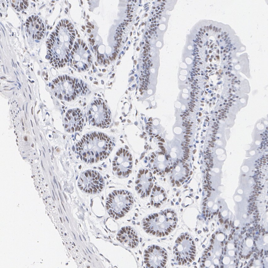
Immunohistochemical analysis of paraffin-embedded rat small intestine tissue with Rabbit anti-KMT3A / SETD2 antibody at 1/200 dilution. The section was pre-treated using heat mediated antigen retrieval with Tris-EDTA buffer (pH 9.0) for 20 minutes. The tissues were blocked in 1% BSA for 20 minutes at room temperature, washed with ddH2O and PBS, and then probed with the primary antibody at 1/200 dilution for 1 hour at room temperature. The detection was performed using an HRP conjugated compact polymer system. DAB was used as the chromogen. Tissues were counterstained with hematoxylin and mounted with DPX.ICC/IF
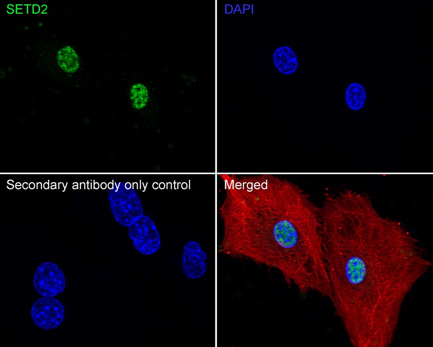
Immunocytochemistry analysis of MEF cells labeling KMT3A / SETD2 with Rabbit anti-KMT3A / SETD2 antibody at 1/100 dilution. Cells were fixed in 4% paraformaldehyde for 15 minutes at room temperature, permeabilized with 0.1% Triton X-100 in PBS for 15 minutes at room temperature, then blocked with 1% BSA in 10% negative goat serum for 1 hour at room temperature. Cells were then incubated with Rabbit anti-KMT3A / SETD2 antibody at 1/100 dilution in 1% BSA in PBST overnight at 4 ℃. Goat Anti-Rabbit IgG H&L (488) was used as the secondary antibody at 1/1,000 dilution. PBS instead of the primary antibody was used as the secondary antibody only control. Nuclear DNA was labelled in blue with DAPI. Beta tubulin (red) was stained at 1/100 dilution overnight at +4℃. Goat Anti-Mouse IgG H&L (594) was used as the secondary antibody at 1/1,000 dilution.IP
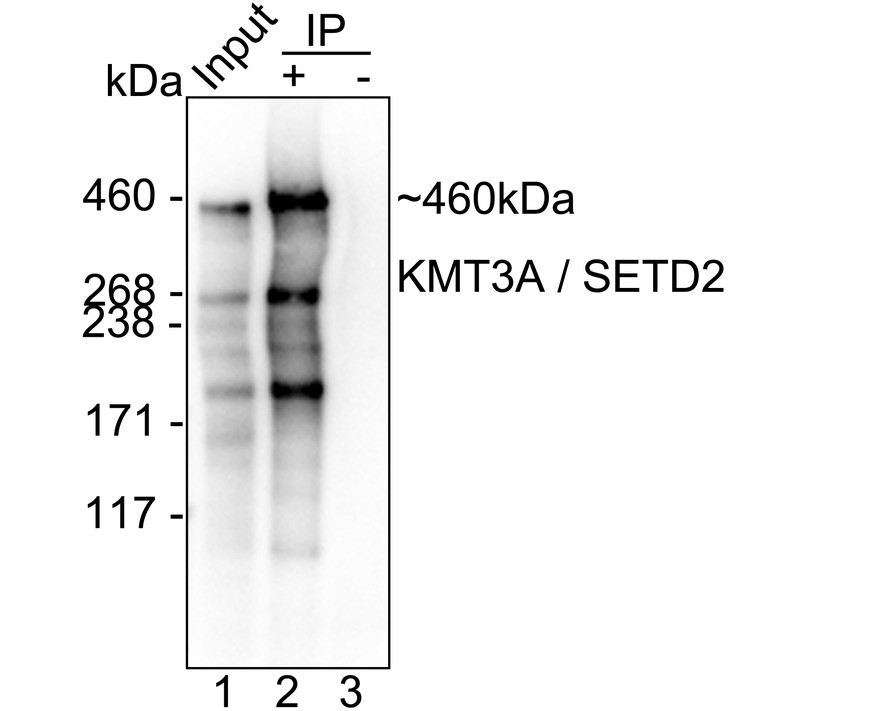
KMT3A / SETD2 was immunoprecipitated from 0.2 mg Daudi cell lysate with KMT3A / SETD2 Recombinant Rabbit Monoclonal Antibodyat 2 µg/10 µl beads. Western blot was performed from the immunoprecipitate using KMT3A / SETD2 Recombinant Rabbit Monoclonal Antibodyat 1/5,000 dilution. HRP Conjugated Anti-Rabbit IgG for IP Nano-secondary antibody at 1/5,000 dilution was used for 1 hour at room temperature. Lane 1: Daudi cell lysate (input). Lane 2: KMT3A / SETD2 Recombinant Rabbit Monoclonal Antibody IP in Daudi cell lysate. Lane 3: Rabbit IgG instead of KMT3A / SETD2 Recombinant Rabbit Monoclonal Antibody in Daudi cell lysate. Blocking/Dilution buffer: primary antibody dilution. Exposure time: 46 seconds.| Product Name | KMT3A / SETD2 Recombinant Rabbit Monoclonal Antibody |
|---|---|
| Antibody Type | Primary Antibodies |
| Immunogen | Recombinant protein within human SETD2 aa 2,051-2,564. |
| Clonality | Monoclonal |
|---|---|
| Isotype | IgG |
| Host Species | Rabbit |
| Tested Applications | ICC/IFIHCIPWB |
| WB:1:5000 IHC:1:200 ICC/IF:1:100 IP:1-2μg/sample |
|
| Species Reactivity | HumanMouseRat |
| Concentration | 1mg/ml |
| Purification | Protein A |
| Gene Symbol | SETD2 |
|---|---|
| Gene Synonyms | LLS HYPB SET2 HIF-1 HIP-1 KMT3A MRD70 RAPAS HBP231 HSPC069 p231HBP |
| Gene Full Name | SET domain containing 2, histone lysine methyltransferase |
| Gene Summary | Huntington's disease (HD), a neurodegenerative disorder characterized by loss of striatal neurons, is caused by an expansion of a polyglutamine tract in the HD protein huntingtin. This gene encodes a protein belonging to a class of huntingtin interacting proteins characterized by WW motifs. This protein is a histone methyltransferase that is specific for lysine-36 of histone H3, and methylation of this residue is associated with active chromatin. This protein also contains a novel transcriptional activation domain and has been found associated with hyperphosphorylated RNA polymerase II. [provided by RefSeq, Aug 2008] |
| Alternative Names | EC 2.1.1.43 antibody FLJ16420 antibody FLJ22472 antibody FLJ23184 antibody FLJ45883 antibody HBP231 antibody HIF 1 antibody HIF-1 antibody HIF1 antibody HIP-1 antibody Histone lysine N methyltransferase SETD2 antibody Histone-lysine N-methyltransferase SETD2 antibody hSET2 antibody HSPC069 antibody Huntingtin interacting protein 1 antibody Huntingtin interacting protein antibody Huntingtin interacting protein B antibody Huntingtin interacting protein HYPB antibody Huntingtin yeast partner B antibody Huntingtin-binding protein 231-KD antibody Huntingtin-interacting protein 1 antibody Huntingtin-interacting protein B antibody HYPB antibody KIAA1732 antibody KMT3A antibody Lysine N methyltransferase 3A antibody Lysine N-methyltransferase 3A antibody p231HBP antibody SET domain containing 2 antibody SET domain-containing protein 2 antibody SET2 antibody SETD2 antibody SETD2_HUMAN antibody |
| Molecular Weight(MW) | 288 kDa(Observed band size: 460 kDa) |
| Cellular Localization | Nucleus, Chromosome. |
WB
Western blot analysis of KMT3A / SETD2 on different lysates with Rabbit anti-KMT3A / SETD2 antibody at 1/5,000 dilution. Lysates/proteins at 20 µg/Lane. Exposure time: 30 seconds; 3-8% SDS-PAGE gel. Proteins were transferred to a PVDF membrane and blocked with 5% NFDM/TBST for 1 hour at room temperature. The primary antibody at 1/5,000 dilution was used in primary antibody dilution at 4℃ overnight. Goat Anti-Rabbit IgG - HRP Secondary Antibody at 1/50,000 dilution was used for 1 hour at room temperature.
IHC
Immunohistochemical analysis of paraffin-embedded human small intestine tissue with Rabbit anti-KMT3A / SETD2 antibody at 1/200 dilution. The section was pre-treated using heat mediated antigen retrieval with Tris-EDTA buffer (pH 9.0) for 20 minutes. The tissues were blocked in 1% BSA for 20 minutes at room temperature, washed with ddH2O and PBS, and then probed with the primary antibody at 1/200 dilution for 1 hour at room temperature. The detection was performed using an HRP conjugated compact polymer system. DAB was used as the chromogen. Tissues were counterstained with hematoxylin and mounted with DPX.
IHC
Immunohistochemical analysis of paraffin-embedded mouse small intestine tissue with Rabbit anti-KMT3A / SETD2 antibody at 1/200 dilution. The section was pre-treated using heat mediated antigen retrieval with Tris-EDTA buffer (pH 9.0) for 20 minutes. The tissues were blocked in 1% BSA for 20 minutes at room temperature, washed with ddH2O and PBS, and then probed with the primary antibody at 1/200 dilution for 1 hour at room temperature. The detection was performed using an HRP conjugated compact polymer system. DAB was used as the chromogen. Tissues were counterstained with hematoxylin and mounted with DPX.
IHC
Immunohistochemical analysis of paraffin-embedded rat small intestine tissue with Rabbit anti-KMT3A / SETD2 antibody at 1/200 dilution. The section was pre-treated using heat mediated antigen retrieval with Tris-EDTA buffer (pH 9.0) for 20 minutes. The tissues were blocked in 1% BSA for 20 minutes at room temperature, washed with ddH2O and PBS, and then probed with the primary antibody at 1/200 dilution for 1 hour at room temperature. The detection was performed using an HRP conjugated compact polymer system. DAB was used as the chromogen. Tissues were counterstained with hematoxylin and mounted with DPX.
ICC/IF
Immunocytochemistry analysis of MEF cells labeling KMT3A / SETD2 with Rabbit anti-KMT3A / SETD2 antibody at 1/100 dilution. Cells were fixed in 4% paraformaldehyde for 15 minutes at room temperature, permeabilized with 0.1% Triton X-100 in PBS for 15 minutes at room temperature, then blocked with 1% BSA in 10% negative goat serum for 1 hour at room temperature. Cells were then incubated with Rabbit anti-KMT3A / SETD2 antibody at 1/100 dilution in 1% BSA in PBST overnight at 4 ℃. Goat Anti-Rabbit IgG H&L (488) was used as the secondary antibody at 1/1,000 dilution. PBS instead of the primary antibody was used as the secondary antibody only control. Nuclear DNA was labelled in blue with DAPI. Beta tubulin (red) was stained at 1/100 dilution overnight at +4℃. Goat Anti-Mouse IgG H&L (594) was used as the secondary antibody at 1/1,000 dilution.
IP
KMT3A / SETD2 was immunoprecipitated from 0.2 mg Daudi cell lysate with KMT3A / SETD2 Recombinant Rabbit Monoclonal Antibodyat 2 µg/10 µl beads. Western blot was performed from the immunoprecipitate using KMT3A / SETD2 Recombinant Rabbit Monoclonal Antibodyat 1/5,000 dilution. HRP Conjugated Anti-Rabbit IgG for IP Nano-secondary antibody at 1/5,000 dilution was used for 1 hour at room temperature. Lane 1: Daudi cell lysate (input). Lane 2: KMT3A / SETD2 Recombinant Rabbit Monoclonal Antibody IP in Daudi cell lysate. Lane 3: Rabbit IgG instead of KMT3A / SETD2 Recombinant Rabbit Monoclonal Antibody in Daudi cell lysate. Blocking/Dilution buffer: primary antibody dilution. Exposure time: 46 seconds.| Application Notes | WB:1:5000 IHC:1:200 ICC/IF:1:100 IP:1-2μg/sample |
|---|
| Form | Liquid |
|---|---|
| Storage Instructions | Shipped at 4℃. Store at +4℃ short term (1-2 weeks). Store at -20℃ long term. |
| Storage Buffer | 1*PBS (pH7.4), 0.1% BSA, 40% Glycerol, 0.2% Proclean 950. |


