ICC/IF
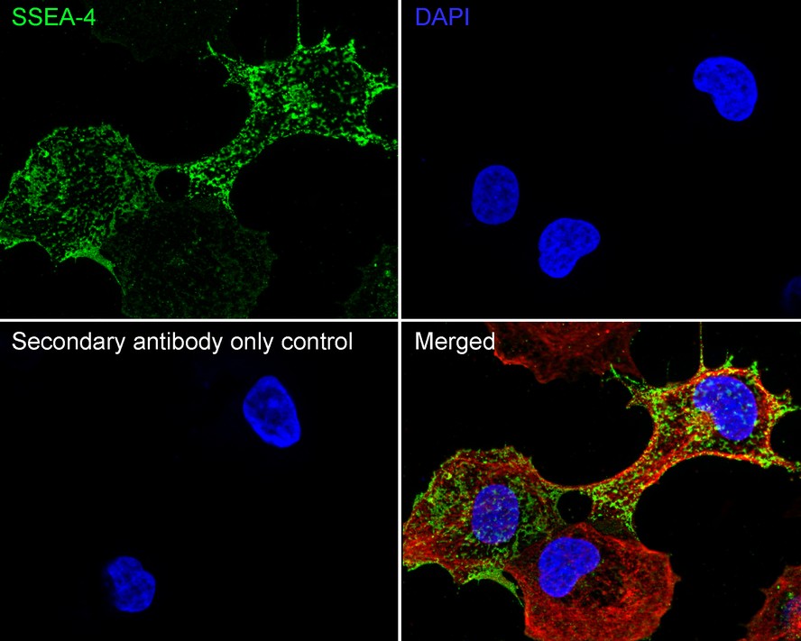
Immunocytochemistry analysis of NTERA-2 cells labeling SSEA4 with Mouse anti-SSEA4 antibody at 1/100 dilution. Cells were fixed in 4% paraformaldehyde for 15 minutes at room temperature, permeabilized with 0.1% Triton X-100 in PBS for 15 minutes at room temperature, then blocked with 1% BSA in 10% negative goat serum for 1 hour at room temperature. Cells were then incubated with Mouse anti-SSEA4 antibody at 1/100 dilution in 1% BSA in PBST overnight at 4 ℃. Goat Anti-Mouse IgG H&L (488) was used as the secondary antibody at 1/1,000 dilution. PBS instead of the primary antibody was used as the secondary antibody only control. Nuclear DNA was labelled in blue with DAPI. beta Tubulin (red) was stained at 1/100 dilution overnight at +4℃. Goat Anti-Rabbit IgG H&L (594) were used as the secondary antibody at 1/1,000 dilution.FC
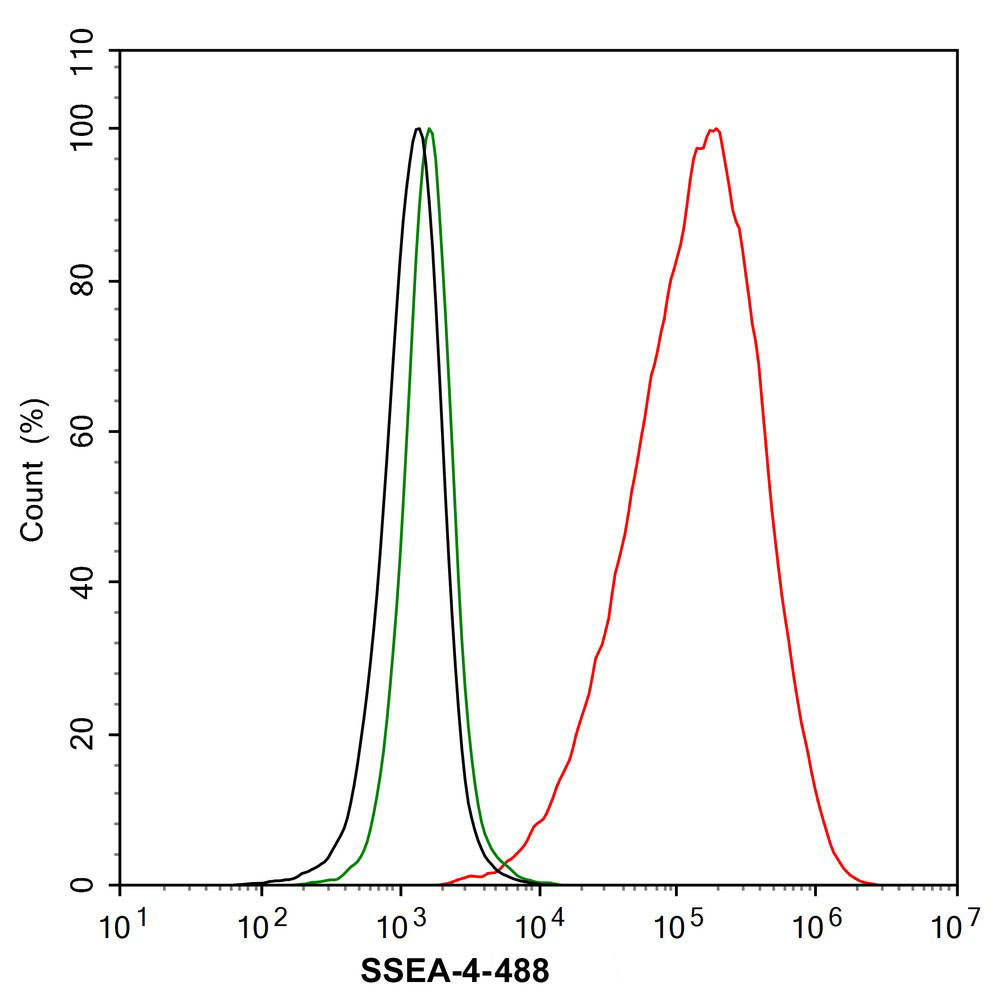
Flow cytometric analysis of NTERA-2 cells labeling SSEA4. Cells were washed twice with cold PBS and resuspend. Then stained with the primary antibody (1/1,000) (red) compared with Mouse IgG Isotype Control (green). After incubation of the primary antibody at +4℃ for an hour, the cells were stained with a 488 conjugate-Goat anti-Mouse IgG Secondary antibody at 1/1,000 dilution for 30 minutes at +4℃. Unlabelled sample was used as a control (cells without incubation with primary antibody; black).| Product Name | SSEA4 Recombinant Mouse Monoclonal Antibody |
|---|---|
| Antibody Type | Primary Antibodies |
| Clonality | Monoclonal |
|---|---|
| Isotype | IgG1 |
| Host Species | Mouse |
| Tested Applications | FCICC/IF |
| ICC/IF:1:100 FC:1:1000 |
|
| Species Reactivity | Human |
| Concentration | 1mg/ml |
| Purification | Protein A |
| Function | SSEA4 (stage-specific embryonic antigen 4) is a glycolipid carbohydrate epitope expressed on the surface of human teratocarcinoma stem cells, human embryonic germ cells, and human embryonic stem cells. Expression of human SSEA4 decreases following differentiation of human embryonal carcinoma cells. Expression of the SSEA4 antigen is absent in murine pluripotent cells, but increases following differentiation. |
|---|
ICC/IF
Immunocytochemistry analysis of NTERA-2 cells labeling SSEA4 with Mouse anti-SSEA4 antibody at 1/100 dilution. Cells were fixed in 4% paraformaldehyde for 15 minutes at room temperature, permeabilized with 0.1% Triton X-100 in PBS for 15 minutes at room temperature, then blocked with 1% BSA in 10% negative goat serum for 1 hour at room temperature. Cells were then incubated with Mouse anti-SSEA4 antibody at 1/100 dilution in 1% BSA in PBST overnight at 4 ℃. Goat Anti-Mouse IgG H&L (488) was used as the secondary antibody at 1/1,000 dilution. PBS instead of the primary antibody was used as the secondary antibody only control. Nuclear DNA was labelled in blue with DAPI. beta Tubulin (red) was stained at 1/100 dilution overnight at +4℃. Goat Anti-Rabbit IgG H&L (594) were used as the secondary antibody at 1/1,000 dilution.
FC
Flow cytometric analysis of NTERA-2 cells labeling SSEA4. Cells were washed twice with cold PBS and resuspend. Then stained with the primary antibody (1/1,000) (red) compared with Mouse IgG Isotype Control (green). After incubation of the primary antibody at +4℃ for an hour, the cells were stained with a 488 conjugate-Goat anti-Mouse IgG Secondary antibody at 1/1,000 dilution for 30 minutes at +4℃. Unlabelled sample was used as a control (cells without incubation with primary antibody; black).| Application Notes | ICC/IF:1:100 FC:1:1000 |
|---|
| Form | Liquid |
|---|---|
| Storage Instructions | Shipped at 4℃. Store at +4℃ short term (1-2 weeks). Store at -20℃ long term. |
| Storage Buffer | 1*PBS (pH7.4), 0.1% BSA, 40% Glycerol, 0.2% Proclean 950. |


