WB
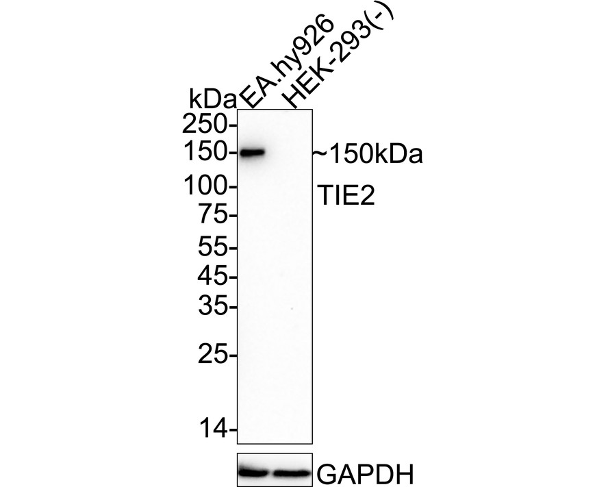
Western blot analysis of TIE2 on different lysates with Rabbit anti-TIE2 antibody at 1/1,000 dilution. Lysates/proteins at 20 µg/Lane. Exposure time: 3 minutes; 4-20% SDS-PAGE gel. Proteins were transferred to a PVDF membrane and blocked with 5% NFDM/TBST for 1 hour at room temperature. The primary antibody at 1/1,000 dilution was used in 5% NFDM/TBST at 4℃ overnight. Goat Anti-Rabbit IgG - HRP Secondary Antibody at 1/50,000 dilution was used for 1 hour at room temperature.FC
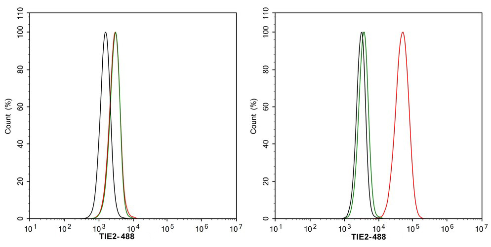
Flow cytometric analysis of HEK-293 (left, negative) and EA.hy926 (right, positive) cells labeling TIE2. Cells were washed twice with cold PBS and resuspend. Then stained with the primary antibody (1μg/mL) (red) compared with Rabbit IgG Isotype Control (green). After incubation of the primary antibody at +4℃ for an hour, the cells were stained with a 488 conjugate-Goat anti-Rabbit IgG Secondary antibody at 1/1,000 dilution for 30 minutes at +4℃. Unlabelled sample was used as a control (cells without incubation with primary antibody; black).| Product Name | TIE2 Recombinant Rabbit Monoclonal Antibody |
|---|---|
| Antibody Type | Primary Antibodies |
| Immunogen | Recombinant protein within human TIE2 aa 1-769. |
| Clonality | Monoclonal |
|---|---|
| Isotype | IgG |
| Host Species | Rabbit |
| Tested Applications | FCWB |
| WB:1:1000 FC:1:1000 |
|
| Species Reactivity | Human |
| Concentration | 1mg/ml |
| Purification | Protein A |
| Gene Symbol | TEK |
|---|---|
| Gene Synonyms | TIE2 VMCM GLC3E TIE-2 VMCM1 CD202B |
| Gene Full Name | TEK receptor tyrosine kinase |
| Gene Summary | This gene encodes a receptor that belongs to the protein tyrosine kinase Tie2 family. The encoded protein possesses a unique extracellular region that contains two immunoglobulin-like domains, three epidermal growth factor (EGF)-like domains and three fibronectin type III repeats. The ligand angiopoietin-1 binds to this receptor and mediates a signaling pathway that functions in embryonic vascular development. Mutations in this gene are associated with inherited venous malformations of the skin and mucous membranes. Alternative splicing results in multiple transcript variants. Additional alternatively spliced transcript variants of this gene have been described, but their full-length nature is not known. [provided by RefSeq, Feb 2014] |
| Alternative Names | Angiopoietin 1 receptor antibody Angiopoietin-1 receptor antibody CD202b antibody CD202b antigen antibody Endothelial tyrosine kinase antibody Endothelium specific receptor tyrosine kinase 2 antibody hTIE 2 antibody hTIE2 antibody Hyk antibody p140 TEK antibody Soluble TIE2 variant 1 antibody Soluble TIE2 variant 2 antibody Tek antibody tek tyrosine kinase antibody TEK tyrosine kinase endothelial antibody tek tyrosine kinase endothelial antibody TIE 2 antibody TIE2 antibody TIE2_HUMAN antibody Tunica interna endothelial cell kinase antibody Tyrosine kinase with Ig and EGF homology domains 2 antibody Tyrosine kinase with Ig and EGF homology domains-2 antibody Tyrosine protein kinase receptor TEK antibody Tyrosine protein kinase receptor TIE 2 antibody Tyrosine-protein kinase receptor TEK antibody Tyrosine-protein kinase receptor TIE-2 antibody Venous malformations multiple cutaneous and mucosal antibody VMCM 1 antibody VMCM antibody VMCM1 antibody |
| Molecular Weight(MW) | 126 kDa(Observed band size: 150 kDa) |
| Cellular Localization | Cell membrane, Cell junction, focal adhesion, Cytoplasm, cytoskeleton, Secreted. |
| SwissProt ID | Q02763 |
|---|
WB
Western blot analysis of TIE2 on different lysates with Rabbit anti-TIE2 antibody at 1/1,000 dilution. Lysates/proteins at 20 µg/Lane. Exposure time: 3 minutes; 4-20% SDS-PAGE gel. Proteins were transferred to a PVDF membrane and blocked with 5% NFDM/TBST for 1 hour at room temperature. The primary antibody at 1/1,000 dilution was used in 5% NFDM/TBST at 4℃ overnight. Goat Anti-Rabbit IgG - HRP Secondary Antibody at 1/50,000 dilution was used for 1 hour at room temperature.
FC
Flow cytometric analysis of HEK-293 (left, negative) and EA.hy926 (right, positive) cells labeling TIE2. Cells were washed twice with cold PBS and resuspend. Then stained with the primary antibody (1μg/mL) (red) compared with Rabbit IgG Isotype Control (green). After incubation of the primary antibody at +4℃ for an hour, the cells were stained with a 488 conjugate-Goat anti-Rabbit IgG Secondary antibody at 1/1,000 dilution for 30 minutes at +4℃. Unlabelled sample was used as a control (cells without incubation with primary antibody; black).| Application Notes | WB:1:1000 FC:1:1000 |
|---|
| Form | Liquid |
|---|---|
| Storage Instructions | Shipped at 4℃. Store at +4℃ short term (1-2 weeks). Store at -20℃ long term. |
| Storage Buffer | PBS (pH7.4), 0.1% BSA, 40% Glycerol. Preservative: 0.05% Sodium Azide. |


