WB
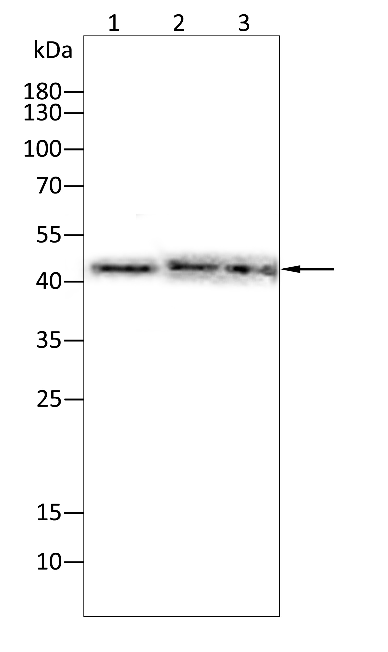
Western blot analysis using ERK1/ 2 mouse monoclonal antibody against Hepg2(1),P19(2),B16(3) cell lysate . 12% SDS-PAGE gel.Sample loading: 20μg /lane. Transfer the proteins onto a PVDF membrane (OM790003), and block it with TBST (OM750016) plus skimmed milk powder(OM750084) for one hour. Dilute the primary antibody with the antibody diluent (OM750012) at a ratio of 1:1000, and incubate it overnight at 4°C. Wash the membrane three times with TBST (OM750016), 5 minutes each time. At room temperature, dilute the secondary antibody, Goat Anti-Mouse IgG (H&L) - HRP(OM644366), at a ratio of 1:20000 and incubate for one hour. Wash the membrane three times with TBST (OM750016) again, 5 minutes each time. Use ECL (OM625701) for luminescence.staining time: 60SICC/IF
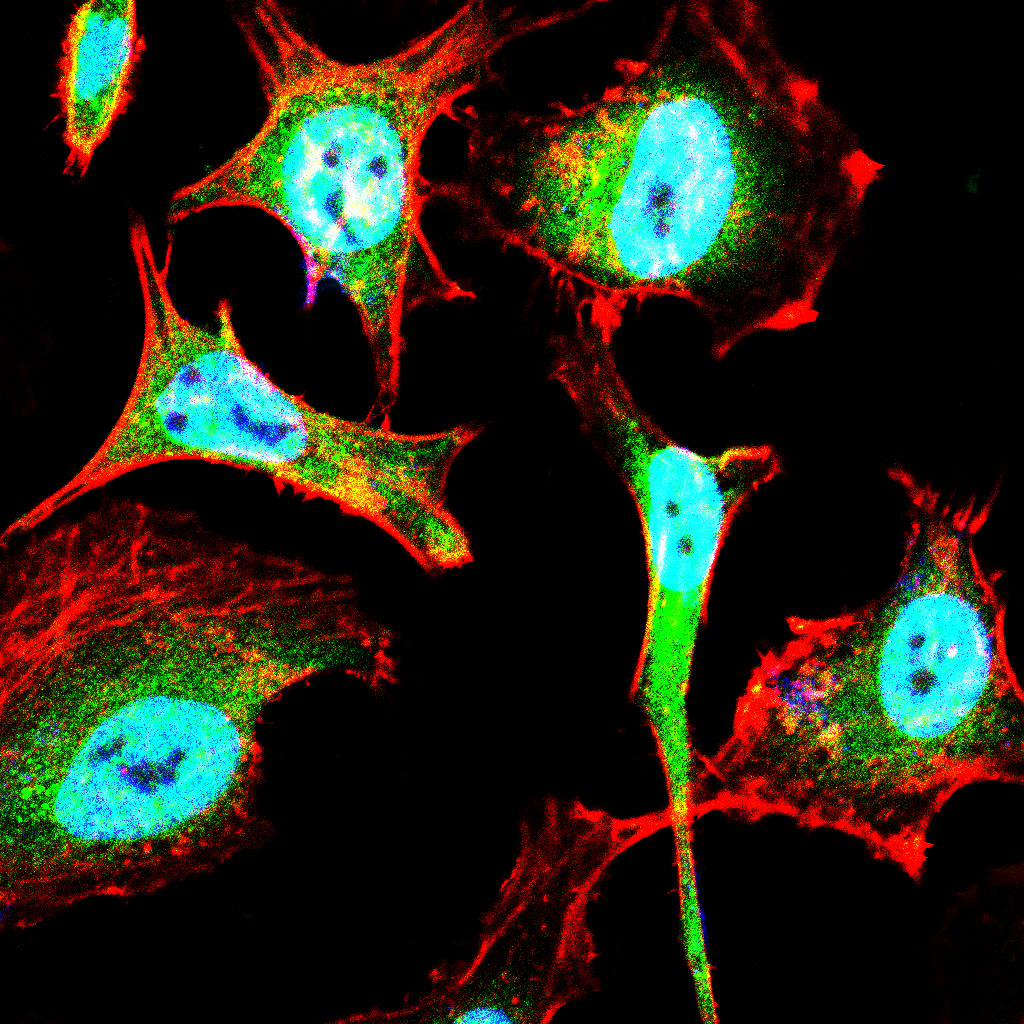
Immunofluorescence analysis of Hela cells using ERK1/2 antibody (green). Blue: DAPI fluorescent DNA dye. Red: Actin filaments have been labeled with Omnimabs® 594-Phalloidin.Cells are fixed in 4% paraformaldehyde at room temperature for 15 minutes. Then, they are permeabilized with a PBS (OM750003) solution containing 0.1% Triton X-100(OM750021) at room temperature for 15 minutes. Subsequently, the cells are blocked with 10% non - immune goat serum(OM760028) at room temperature for 1 hour.The cells are incubated overnight at 4°C with the primary antibody diluted 1:100 in PBS. The secondary antibody, Omnimabs® 488 Goat Ant-Mouse IgG(H&L) (Green,OM643485), is diluted at a ratio of 1:400 and incubated with the cells for 1 hour.Nuclear DNA is labeled with DAPI (Blue,OM643160). F-actin is stained with Omnimabs® 594-Phalloidin (Red,OM750007) diluted 1:100 for 30 minutes.| Product Name | Anti-ERK1/2 antibody |
|---|---|
| Antibody Type | Primary Antibodies |
| Immunogen | Recombinant protein |
| Clonality | Monoclonal |
|---|---|
| Isotype | IgG |
| Host Species | Mouse |
| Tested Applications | ICC/IFWB |
| WB:1:1000 ICC/IF:1:100-1:500 ELISA:1:1000-1:10000 |
|
| Species Reactivity | HumanMouseRat |
| Concentration | 1mg/ml |
| Purification | Affinity purified |
| Gene Symbol | MAPK1/3 |
|---|---|
| Gene Synonyms | ERK p38 p40 p41 ERK2 ERT1 NS13 ERK-2 MAPK2 PRKM1 PRKM2 P42MAPK p41mapk p42-MAPK |
| Gene Full Name | mitogen-activated protein kinase 1/3 |
| Gene Summary | This gene encodes a member of the MAP kinase family. MAP kinases, also known as extracellular signal-regulated kinases (ERKs), act as an integration point for multiple biochemical signals, and are involved in a wide variety of cellular processes such as proliferation, differentiation, transcription regulation and development. The activation of this kinase requires its phosphorylation by upstream kinases. Upon activation, this kinase translocates to the nucleus of the stimulated cells, where it phosphorylates nuclear targets. One study also suggests that this protein acts as a transcriptional repressor independent of its kinase activity. The encoded protein has been identified as a moonlighting protein based on its ability to perform mechanistically distinct functions. Two alternatively spliced transcript variants encoding the same protein, but differing in the UTRs, have been reported for this gene. [provided by RefSeq, Jan 2014] |
| Alternative Names | ERK 1 antibody ERK 2 antibody ERK-1 antibody ERK-2 antibody ERK1 antibody erk1/2 antibody ERK2 antibody ERT1 antibody ERT2 antibody Extracellular signal regulated kinase 1 antibody |
| Molecular Weight(MW) | 44/42kDa |
| Cellular Localization | Cytoplasm Nucleus Membrane, Cell junction, focal adhesion |
WB
Western blot analysis using ERK1/ 2 mouse monoclonal antibody against Hepg2(1),P19(2),B16(3) cell lysate . 12% SDS-PAGE gel.Sample loading: 20μg /lane. Transfer the proteins onto a PVDF membrane (OM790003), and block it with TBST (OM750016) plus skimmed milk powder(OM750084) for one hour. Dilute the primary antibody with the antibody diluent (OM750012) at a ratio of 1:1000, and incubate it overnight at 4°C. Wash the membrane three times with TBST (OM750016), 5 minutes each time. At room temperature, dilute the secondary antibody, Goat Anti-Mouse IgG (H&L) - HRP(OM644366), at a ratio of 1:20000 and incubate for one hour. Wash the membrane three times with TBST (OM750016) again, 5 minutes each time. Use ECL (OM625701) for luminescence.staining time: 60S
ICC/IF
Immunofluorescence analysis of Hela cells using ERK1/2 antibody (green). Blue: DAPI fluorescent DNA dye. Red: Actin filaments have been labeled with Omnimabs® 594-Phalloidin.Cells are fixed in 4% paraformaldehyde at room temperature for 15 minutes. Then, they are permeabilized with a PBS (OM750003) solution containing 0.1% Triton X-100(OM750021) at room temperature for 15 minutes. Subsequently, the cells are blocked with 10% non - immune goat serum(OM760028) at room temperature for 1 hour.The cells are incubated overnight at 4°C with the primary antibody diluted 1:100 in PBS. The secondary antibody, Omnimabs® 488 Goat Ant-Mouse IgG(H&L) (Green,OM643485), is diluted at a ratio of 1:400 and incubated with the cells for 1 hour.Nuclear DNA is labeled with DAPI (Blue,OM643160). F-actin is stained with Omnimabs® 594-Phalloidin (Red,OM750007) diluted 1:100 for 30 minutes.| Application Notes | WB:1:1000 ICC/IF:1:100-1:500 ELISA:1:1000-1:10000 |
|---|
| Form | Liquid |
|---|---|
| Storage Instructions | Shipped at 4°C. Store at +4°C short term (1-2 weeks). Store at -20°C long term. Avoid freeze / thaw cycle. |
| Storage Buffer | Purified antibody in PBS with 0.05% sodium azide,40% Glycerol. |


