ELISA
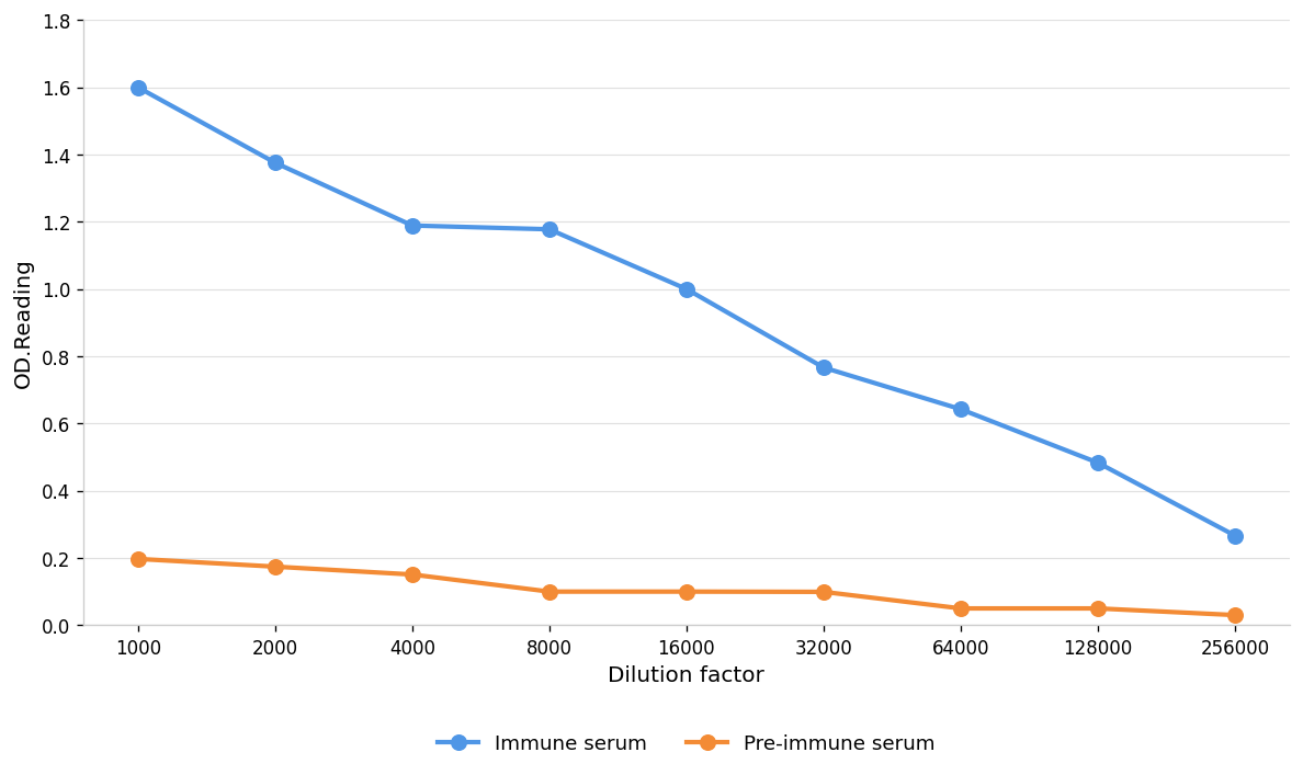
Coat the plate with immunizing antigen.Blue line: Immune serum; Orange line: Pre-immune serum.WB
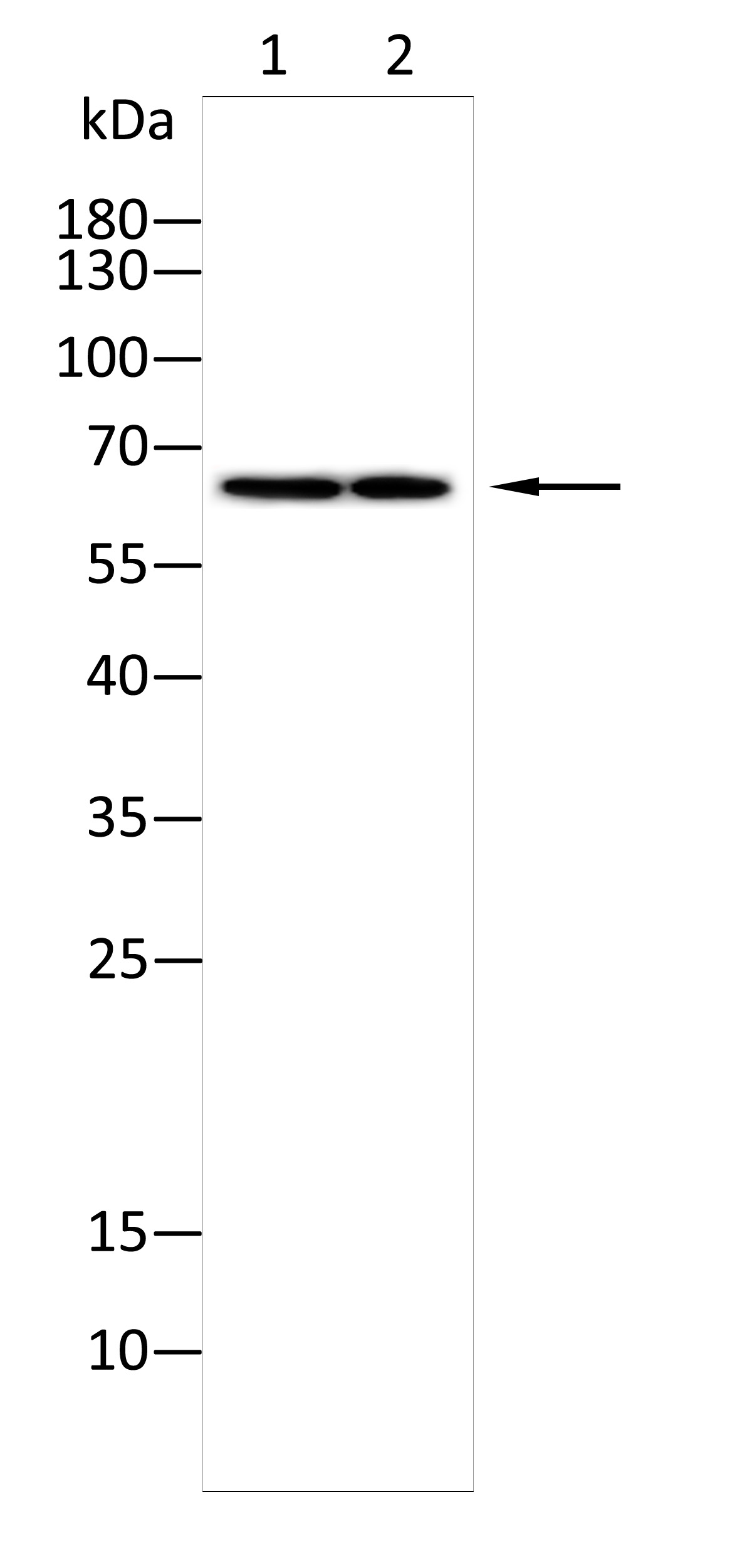
Western blot analysis using SPAM1 antibody against K562(1),Hepg2(2) cell . 12% SDS-PAGE gel.Sample loading: 20μg /lane. Transfer the proteins onto a PVDF membrane (OM790003), and block it with TBST (OM750016) plus skimmed milk powder(OM750084) for one hour. Dilute the primary antibody with the antibody diluent (OM750012) at a ratio of 1:1000, and incubate it overnight at 4°C. Wash the membrane three times with TBST (OM750016), 5 minutes each time. At room temperature, dilute the secondary antibody, Goat Anti-Rabbit IgG(H&L)-HRP (OM643487), at a ratio of 1:20000 and incubate for one hour. Wash the membrane three times with TBST (OM750016) again, 5 minutes each time. Use ECL (OM625701) for luminescence.staining time: 60S.| Product Name | Anti-SPAM1 antibody |
|---|---|
| Antibody Type | Primary Antibodies |
| Immunogen | Polypeptide |
| Clonality | Polyclonal |
|---|---|
| Isotype | IgG |
| Host Species | Rabbit |
| Tested Applications | ELISAWB |
| WB:1:1000-1:2000 ELISA:1:1000-1:10000 |
|
| Species Reactivity | HumanMouseRat |
| Concentration | 1mg/ml |
| Purification | Protein A |
| Gene Symbol | SPAM1 |
|---|---|
| Gene Synonyms | HEL-S-96n HYA1 HYAL5 PH-20 PH20 SPAG15 Spam |
| Gene Full Name | sperm adhesion molecule 1 |
| Gene Summary | Hyaluronidase degrades hyaluronic acid, a major structural proteoglycan found in extracellular matrices and basement membranes. Six members of the hyaluronidase family are clustered into two tightly linked groups on chromosome 3p21.3 and 7q31.3. This gene was previously referred to as HYAL1 and HYA1 and has since been assigned the official symbol SPAM1; another family member on chromosome 3p21.3 has been assigned HYAL1. This gene encodes a GPI-anchored enzyme located on the human sperm surface and inner acrosomal membrane. This multifunctional protein is a hyaluronidase that enables sperm to penetrate through the hyaluronic acid-rich cumulus cell layer surrounding the oocyte, a receptor that plays a role in hyaluronic acid induced cell signaling, and a receptor that is involved in sperm-zona pellucida adhesion. Abnormal expression of this gene in tumors has implicated this protein in degradation of basement membranes leading to tumor invasion and metastasis. Multiple transcript variants encoding different isoforms have been found for this gene. [provided by RefSeq, Mar 2010] |
| Alternative Names | 2B1 antigen epididymis secretory sperm binding protein Li 96n hyal-PH20 hyaluronoglucosaminidase PH-20 MGC26532 PH-20 hyaluronidase sperm adhesion molecule 1 (PH-20 hyaluronidase zona pellucida binding) sperm surface antigen 2B1 sperm surface protein PH-20 |
| Molecular Weight(MW) | 57kDa(observed:62kDa) |
| Source | Rabbit |
| Cellular Localization | Cell membrane;Membrane |
| SwissProt ID | P38567 |
|---|
ELISA
Coat the plate with immunizing antigen.Blue line: Immune serum; Orange line: Pre-immune serum.
WB
Western blot analysis using SPAM1 antibody against K562(1),Hepg2(2) cell . 12% SDS-PAGE gel.Sample loading: 20μg /lane. Transfer the proteins onto a PVDF membrane (OM790003), and block it with TBST (OM750016) plus skimmed milk powder(OM750084) for one hour. Dilute the primary antibody with the antibody diluent (OM750012) at a ratio of 1:1000, and incubate it overnight at 4°C. Wash the membrane three times with TBST (OM750016), 5 minutes each time. At room temperature, dilute the secondary antibody, Goat Anti-Rabbit IgG(H&L)-HRP (OM643487), at a ratio of 1:20000 and incubate for one hour. Wash the membrane three times with TBST (OM750016) again, 5 minutes each time. Use ECL (OM625701) for luminescence.staining time: 60S.| Application Notes | WB:1:1000-1:2000 ELISA:1:1000-1:10000 |
|---|
| Form | Liquid |
|---|---|
| Storage Instructions | Shipped at 4°C. Store at +4°C short term (1-2 weeks). Store at -20°C long term. Avoid freeze / thaw cycle. |
| Storage Buffer | Purified antibody in PBS with 0.05% PC300,40% Glycerol. |


