WB
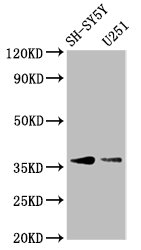
Positive WB detected in: SH-SY5Y whole cell lysate, U251 whole cell lysate All lanes: BARHL1 antibody at 3.4µg/ml Secondary Goat polyclonal to rabbit IgG at 1/50000 dilutionIHC
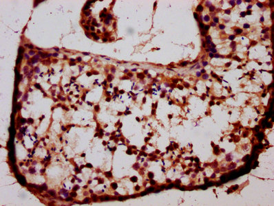
IHC image of BARHL1 Antibody diluted at 1:400 and staining in paraffin-embedded human testis tissue performed on a Leica BondTM system. After dewaxing and hydration, antigen retrieval was mediated by high pressure in a citrate buffer (pH 6.0). Section was blocked with 10% normal goat serum 30min at RT. Then primary antibody (1% BSA) was incubated at 4°C overnight. The primary is detected by a biotinylated secondary antibody and visualized using an HRP conjugated SP system.| Product Name | BARHL1 Antibody |
|---|---|
| Antibody Type | Primary Antibodies |
| Immunogen | Recombinant Human BarH-like 1 homeobox protein (38-176AA) |
| Clonality | Polyclonal |
|---|---|
| Isotype | IgG |
| Host Species | Rabbit |
| Tested Applications | IHCWB |
| WB:1:500-1:5000 IHC:1:200-1:500 |
|
| Species Reactivity | Human |
| Concentration | 1mg/ml |
| Purification | Protein G |
| Gene Symbol | BARHL1 |
|---|---|
| Gene Synonyms | Eukaryota Metazoa Chordata Craniata Vertebrata Euteleostomi Mammalia Eutheria Euarchontoglires Primates Haplorrhini Catarrhini Hominidae Homo |
| Gene Full Name | BarH like homeobox 1 |
| Gene Summary | Enables sequence-specific double-stranded DNA binding activity. Predicted to be involved in regulation of transcription by RNA polymerase II. Predicted to act upstream of or within several processes, including negative regulation of outer hair cell apoptotic process; nervous system development; and sensory perception of sound. Predicted to be located in chromatin. Predicted to be active in nucleus. Biomarker of Alzheimer's disease; high grade glioma; and triple-receptor negative breast cancer. [provided by Alliance of Genome Resources, Jul 2025] |
| Alternative Names | BarH (Drosophila) like 1 antibody BARH L1 antibody BarH like 1 antibody BarH like 1 homeobox protein antibody BarH like homeobox 1 antibody BarH-like 1 homeobox protein antibody BARH1_HUMAN antibody BARHL 1 antibody BARHL1 antibody FKSG 31 antibody FKSG31 antibody |
| Molecular Weight(MW) | 36 kDa |
| Cellular Localization | Nucleus. |
WB
Positive WB detected in: SH-SY5Y whole cell lysate, U251 whole cell lysate All lanes: BARHL1 antibody at 3.4µg/ml Secondary Goat polyclonal to rabbit IgG at 1/50000 dilution
IHC
IHC image of BARHL1 Antibody diluted at 1:400 and staining in paraffin-embedded human testis tissue performed on a Leica BondTM system. After dewaxing and hydration, antigen retrieval was mediated by high pressure in a citrate buffer (pH 6.0). Section was blocked with 10% normal goat serum 30min at RT. Then primary antibody (1% BSA) was incubated at 4°C overnight. The primary is detected by a biotinylated secondary antibody and visualized using an HRP conjugated SP system.| Application Notes | WB:1:500-1:5000 IHC:1:200-1:500 |
|---|
| Form | Liquid |
|---|---|
| Storage Instructions | Upon receipt, store at -20°C or -80°C. Avoid repeated freeze. |
| Storage Buffer | 0.03% Proclin 300 Constituents: 50% Glycerol, 0.01M PBS, PH 7.4 |


