WB
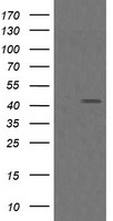
HEK293T cells were transfected with the pCMV6-ENTRY control or pCMV6-ENTRY MAP2K1 cDNA for 48 hrs and lysed. Equivalent amounts of cell lysates (5 ug per lane) were separated by SDS-PAGE and immunoblotted with anti-MAP2K1. Positive lysates LY400974 (100ug) and LC400974 (20ug) can be purchased separately from OriGene.WB
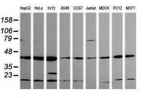
Western blot analysis of extracts (35ug) from 9 different cell lines by usin g anti-MAP2K1 monoclonal antibody (HepG2: human; HeLa: human; SVT2: mouse; A549: human; COS7: monkey; Jurkat: human; MDCK: canine; PC12: rat; MCF7: human).WB
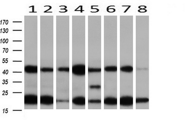
Western blot analysis of extracts (10ug) from 8 Human tissue by using anti-MAP2K1 monoclonal antibody at 1:200 (1: Testis; 2: Uterus; 3: Breast; 4: Brain; 5: Liver; 6: Ovary; 7: Thyroid gland; 8: Colon).IHC
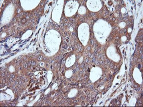
Immunohistochemical staining of paraffin-embedded Adenocarcinoma of Human breast tissue using anti-MAP2K1 mouse monoclonal antibody. Heat-induced epitope retrieval by EDTA solution buffer pH 8.0 at 120°C for 3 min.IHC
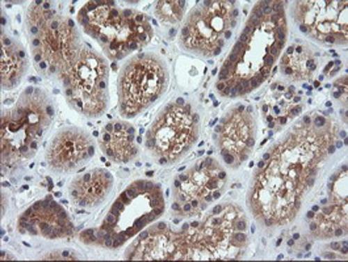
Immunohistochemical staining of paraffin-embedded Human Kidney tissue within the normal limits using anti-MAP2K1 mouse monoclonal antibody. Heat-induced epitope retrieval by EDTA solution buffer pH 8.0 at 120°C for 3 min.IHC
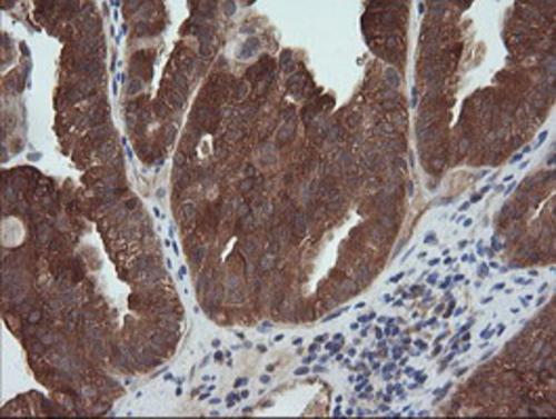
Immunohistochemical staining of paraffin-embedded Adenocarcinoma of Human ovary tissue using anti-MAP2K1 mouse monoclonal antibody. Heat-induced epitope retrieval by EDTA solution buffer pH 8.0 at 120°C for 3 min.IHC
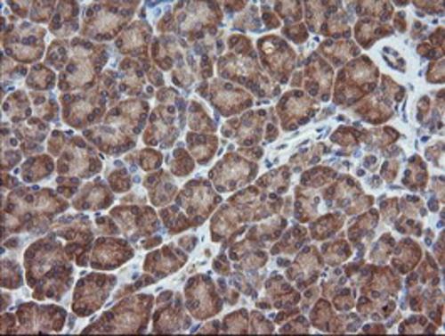
Immunohistochemical staining of paraffin-embedded Human pancreas tissue within the normal limits using anti-MAP2K1 mouse monoclonal antibody. Heat-induced epitope retrieval by EDTA solution buffer pH 8.0 at 120°C for 3 min.ICC/IF
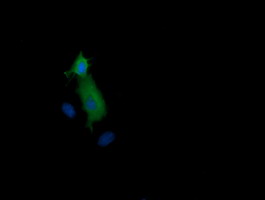
Anti-MAP2K1 mouse monoclonal antibody immunofluorescent staining of COS7 cells transiently transfected by pCMV6-ENTRY MAP2K1.| Product Name | MEK1 (MAP2K1) Mouse Monoclonal Antibody |
|---|---|
| Antibody Type | Primary Antibodies |
| Immunogen | Full length human recombinant protein of human MAP2K1(NP_002746) produced in HEK293T cell. |
| Clonality | monoclonal |
|---|---|
| Isotype | IgG1 |
| Host Species | Mouse |
| Tested Applications | ICC/IFIHCWB |
| WB:1:200-2000 IHC:1:150 ICC/IF:1:100 |
|
| Species Reactivity | Mouse |
| Concentration | 1mg/ml |
| Purification | Protein A/G |
| Gene Symbol | Map2k1 |
|---|---|
| Gene Synonyms | Mek1 MEKK1 MAPKK1 Prkmk1 |
| Gene Full Name | mitogen-activated protein kinase kinase 1 |
| Gene Summary | Enables MAP kinase kinase activity and protein serine/threonine kinase activity. Involved in MAPK cascade; cell surface receptor protein tyrosine kinase signaling pathway; and nervous system development. Acts upstream of with a positive effect on type B pancreatic cell proliferation. Acts upstream of or within several processes, including central nervous system development; regulation of neuron projection development; and respiratory system development. Predicted to be located in several cellular components, including dendrite cytoplasm; perikaryon; and perinuclear region of cytoplasm. Predicted to be active in glutamatergic synapse and postsynaptic density. Is expressed in several structures, including alimentary system; blastocyst; brain; genitourinary system; and sensory organ. Used to study cardiofaciocutaneous syndrome. Human ortholog(s) of this gene implicated in carcinoma (multiple); cardiofaciocutaneous syndrome 3; high grade glioma; and melorheostosis. Orthologous to human MAP2K1 (mitogen-activated protein kinase kinase 1). [provided by Alliance of Genome Resources, Apr 2025] |
| Alternative Names | CFC3 MAPKK1 MEK1 MKK1 PRKMK1 |
| Molecular Weight(MW) | 43.3 kDa |
| Cellular Localization | Cytoplasm,Cytoskeleton,Membrane,Nucleus. |
WB
HEK293T cells were transfected with the pCMV6-ENTRY control or pCMV6-ENTRY MAP2K1 cDNA for 48 hrs and lysed. Equivalent amounts of cell lysates (5 ug per lane) were separated by SDS-PAGE and immunoblotted with anti-MAP2K1. Positive lysates LY400974 (100ug) and LC400974 (20ug) can be purchased separately from OriGene.
WB
Western blot analysis of extracts (35ug) from 9 different cell lines by usin g anti-MAP2K1 monoclonal antibody (HepG2: human; HeLa: human; SVT2: mouse; A549: human; COS7: monkey; Jurkat: human; MDCK: canine; PC12: rat; MCF7: human).
WB
Western blot analysis of extracts (10ug) from 8 Human tissue by using anti-MAP2K1 monoclonal antibody at 1:200 (1: Testis; 2: Uterus; 3: Breast; 4: Brain; 5: Liver; 6: Ovary; 7: Thyroid gland; 8: Colon).
IHC
Immunohistochemical staining of paraffin-embedded Adenocarcinoma of Human breast tissue using anti-MAP2K1 mouse monoclonal antibody. Heat-induced epitope retrieval by EDTA solution buffer pH 8.0 at 120°C for 3 min.
IHC
Immunohistochemical staining of paraffin-embedded Human Kidney tissue within the normal limits using anti-MAP2K1 mouse monoclonal antibody. Heat-induced epitope retrieval by EDTA solution buffer pH 8.0 at 120°C for 3 min.
IHC
Immunohistochemical staining of paraffin-embedded Adenocarcinoma of Human ovary tissue using anti-MAP2K1 mouse monoclonal antibody. Heat-induced epitope retrieval by EDTA solution buffer pH 8.0 at 120°C for 3 min.
IHC
Immunohistochemical staining of paraffin-embedded Human pancreas tissue within the normal limits using anti-MAP2K1 mouse monoclonal antibody. Heat-induced epitope retrieval by EDTA solution buffer pH 8.0 at 120°C for 3 min.
ICC/IF
Anti-MAP2K1 mouse monoclonal antibody immunofluorescent staining of COS7 cells transiently transfected by pCMV6-ENTRY MAP2K1.| Application Notes | WB:1:200-2000 IHC:1:150 ICC/IF:1:100 |
|---|
| Form | Liquid |
|---|---|
| Storage Instructions | Store at -20°C as received. |
| Storage Buffer | PBS (pH 7.3) containing 1% BSA, 50% glycerol and 0.02% sodium azide. |


