ICC/IF
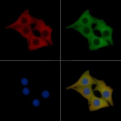
PKD1/PKC mu Mouse Monoclonal Antibody staining Hela cells by IF/ICC. The samples were fixed with PFA and permeabilized in 0.1% Triton X-100, then blocked in 10% serum for 45 minutes at 25°C. Samples were then incubated with primary Ab and rabbit anti-beta tubulin Ab for 1 hour at 37°C. An AlexaFluor594 conjugated goat anti-mouse IgG Ab(Red) and an AlexaFluor488 conjugated goat anti-rabbit IgG Ab(Green) were used as the secondary antibody. The nuclear counter stain is DAPI (blue).WB
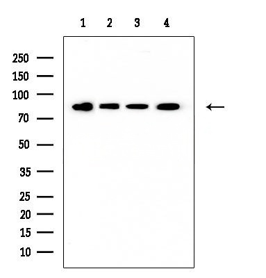
Western blot analysis of extracts from various samples, using PKD1/PKC mu Mouse Monoclonal Antibody at 1/1000 dilution. Lane 1: Rat liver. Lane 2: Colo205 cells. Lane 3: Ec304 cells. Lane 4: Huvec cells. Observed bands:85kD. Exposure for 10s with ECL Kit.WB
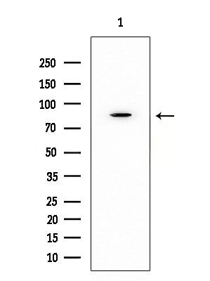
Western blot analysis of extracts from Mouse testis, using PKD1/PKC mu Mouse Monoclonal Antibody at 1/1000 dilution. Observed bands:85kD. Exposure for 10s with ECL Kit.| Product Name | PKD1/PKC mu Mouse Monoclonal Antibody |
|---|---|
| Antibody Type | Primary Antibodies |
| Immunogen | A synthesized peptide derived from human PKD1/PKC mu. |
| Clonality | Monoclonal |
|---|---|
| Isotype | IgG |
| Host Species | Mouse |
| Tested Applications | ICC/IFWB |
| WB:1:500-1:3000 ICC/IF:1:100-1:500 |
|
| Species Reactivity | HumanMouseRat |
| Concentration | 1mg/ml |
| Purification | Affinity purified |
| Gene Symbol | PRKD1 |
|---|---|
| Gene Synonyms | PKD PKCM PKD1 CHDED PRKCM PKC-MU |
| Gene Full Name | protein kinase D1 |
| Gene Summary | The protein encoded by this gene is a serine/threonine protein kinase involved in many cellular processes, including Golgi body membrane integrity and transport, cell migration and differentiation, MAPK8/JNK1 and Ras pathway signaling, MAPK1/3 (ERK1/2) pathway signaling, cell survival, and regulation of cell shape and adhesion. [provided by RefSeq, Jan 2017] |
| Alternative Names | KPCD1_HUMAN nPKC D1 nPKC mu nPKC-D1 nPKC-mu nPKCD1 nPKCmu PKC PKC MU PKCM PKCmu PKD 1 PKD PKD1 PRKCM PRKD 1 Prkd1 Protein kinase C mu Protein kinase C mu type Protein kinase D Protein kinase D1 Serine/threonine protein kinase D1 Serine/threonine-protein kinase D1 |
| Molecular Weight(MW) | 102kD(Calculated). |
| Cellular Localization | Cytoplasm. Cell membrane. Golgi apparatus>trans-Golgi network. |
ICC/IF
PKD1/PKC mu Mouse Monoclonal Antibody staining Hela cells by IF/ICC. The samples were fixed with PFA and permeabilized in 0.1% Triton X-100, then blocked in 10% serum for 45 minutes at 25°C. Samples were then incubated with primary Ab and rabbit anti-beta tubulin Ab for 1 hour at 37°C. An AlexaFluor594 conjugated goat anti-mouse IgG Ab(Red) and an AlexaFluor488 conjugated goat anti-rabbit IgG Ab(Green) were used as the secondary antibody. The nuclear counter stain is DAPI (blue).
WB
Western blot analysis of extracts from various samples, using PKD1/PKC mu Mouse Monoclonal Antibody at 1/1000 dilution. Lane 1: Rat liver. Lane 2: Colo205 cells. Lane 3: Ec304 cells. Lane 4: Huvec cells. Observed bands:85kD. Exposure for 10s with ECL Kit.
WB
Western blot analysis of extracts from Mouse testis, using PKD1/PKC mu Mouse Monoclonal Antibody at 1/1000 dilution. Observed bands:85kD. Exposure for 10s with ECL Kit.| Application Notes | WB:1:500-1:3000 ICC/IF:1:100-1:500 |
|---|
| Form | Liquid |
|---|---|
| Storage Instructions | Store at -20 °C. Stable for 12 months from date of receipt. |
| Storage Buffer | Mouse IgG1 in phosphate buffered saline (without Mg2+ and Ca2+), pH 7.4, 150mM NaCl, 0.02% sodium azide and 50% glycerol. |


