WB
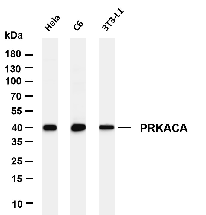
Various whole cell lysates were separated by 4-20% SDS-PAGE, and the membrane was blotted with anti-PRKACA antibody. The HRP-conjugated Goat anti-Rabbit IgG (H + L) antibody was used to detect the antibody. Lane 1: Hela, Lane 2: C6, Lane 3: 3T3-L1 ,IHC
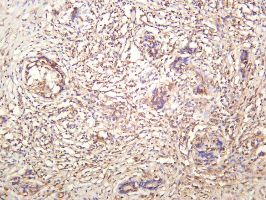
Human thyroid carcinoma was stained with anti-PRKACA rabbit antibodyIHC
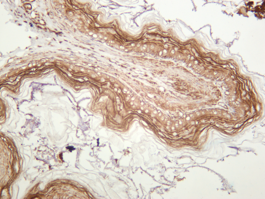
Rat stomach was stained with anti-PRKACA rabbit antibodyICC/IF
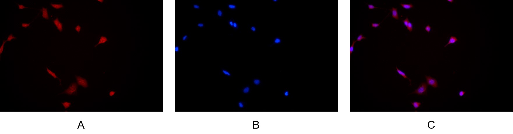
Immunofluorescence analysis of HEK293. Picture A: PRKACA antibody (red). Picture B: DAPI (blue). Picture C: Merge of A+B| Product Name | PRKACA Rabbit mAb |
|---|---|
| Antibody Type | Primary Antibodies |
| Immunogen | The specific immunogen used to produce this antibody is proprietary information. |
| Modification | p-Ser614/Ser619 |
| Clonality | Monoclonal |
|---|---|
| Isotype | IgG,K |
| Host Species | Rabbit |
| Tested Applications | ICC/IFIHCWB |
| WB:1:1000-1:5000 IHC:1:2000-1:10000 ICC/IF:1:200-1:1000 |
|
| Species Reactivity | HumanMouseRat |
| Concentration | 1mg/ml |
| Purification | Affinity purified |
| Gene Symbol | PRKACA |
|---|---|
| Gene Synonyms | CAFD1 PKACA PPNAD4 |
| Gene Full Name | protein kinase cAMP-activated catalytic subunit alpha |
| Gene Summary | This gene encodes one of the catalytic subunits of protein kinase A, which exists as a tetrameric holoenzyme with two regulatory subunits and two catalytic subunits, in its inactive form. cAMP causes the dissociation of the inactive holoenzyme into a dimer of regulatory subunits bound to four cAMP and two free monomeric catalytic subunits. Four different regulatory subunits and three catalytic subunits have been identified in humans. cAMP-dependent phosphorylation of proteins by protein kinase A is important to many cellular processes, including differentiation, proliferation, and apoptosis. Constitutive activation of this gene caused either by somatic mutations, or genomic duplications of regions that include this gene, have been associated with hyperplasias and adenomas of the adrenal cortex and are linked to corticotropin-independent Cushing's syndrome. Alternative splicing results in multiple transcript variants encoding different isoforms. Tissue-specific isoforms that differ at the N-terminus have been described, and these isoforms may differ in the post-translational modifications that occur at the N-terminus of some isoforms. [provided by RefSeq, Jan 2015] |
| Alternative Names | PRKACA PKACA cAMP-dependent protein kinase catalytic subunit alpha PKA C-alpha |
| Molecular Weight(MW) | 40 kDa |
| Cellular Localization | Cytoplasm , Membrane |
WB
Various whole cell lysates were separated by 4-20% SDS-PAGE, and the membrane was blotted with anti-PRKACA antibody. The HRP-conjugated Goat anti-Rabbit IgG (H + L) antibody was used to detect the antibody. Lane 1: Hela, Lane 2: C6, Lane 3: 3T3-L1 ,
IHC
Human thyroid carcinoma was stained with anti-PRKACA rabbit antibody
IHC
Rat stomach was stained with anti-PRKACA rabbit antibody
ICC/IF
Immunofluorescence analysis of HEK293. Picture A: PRKACA antibody (red). Picture B: DAPI (blue). Picture C: Merge of A+B| Application Notes | WB:1:1000-1:5000 IHC:1:2000-1:10000 ICC/IF:1:200-1:1000 |
|---|
| Form | Liquid |
|---|---|
| Storage Instructions | -15°C to -25°C/1 year(Do not lower than -25°C) |
| Storage Buffer | PBS, 50% glycerol, 0.05% Proclin 300, 0.05%BSA |


