WB
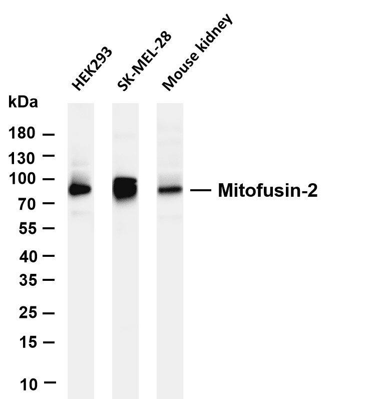
Various whole cell lysates were separated by 4-20% SDS-PAGE, and the membrane was blotted with anti-Mitofusin-2 antibody. The HRP-conjugated Goat anti-Rabbit IgG (H + L) antibody was used to detect the antibody. Lane 1: HEK293, Lane 2: SK-MEL-28, Lane 3: Mouse kidney.IHC
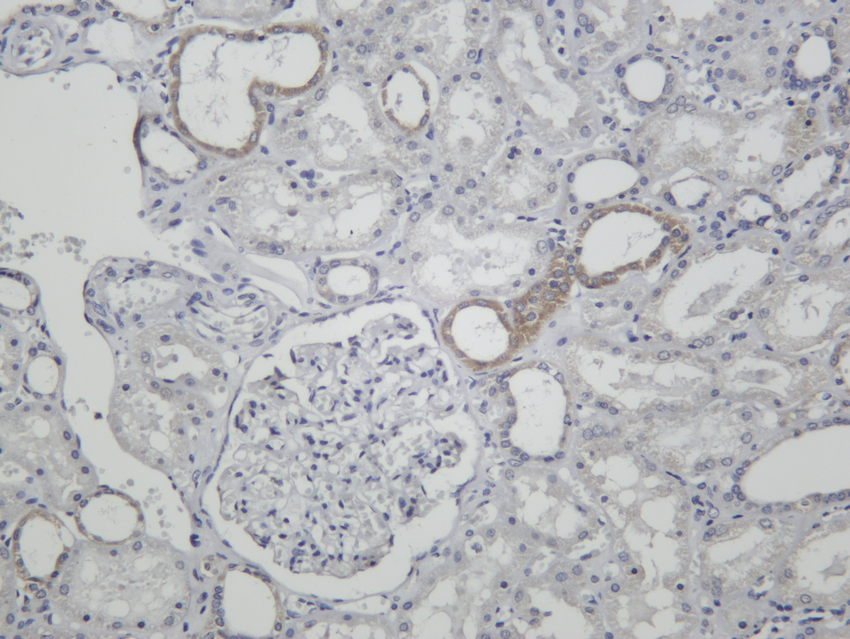
Human kidney was stained with anti-Mitofusin-2 Rabbit antibodyICC/IF
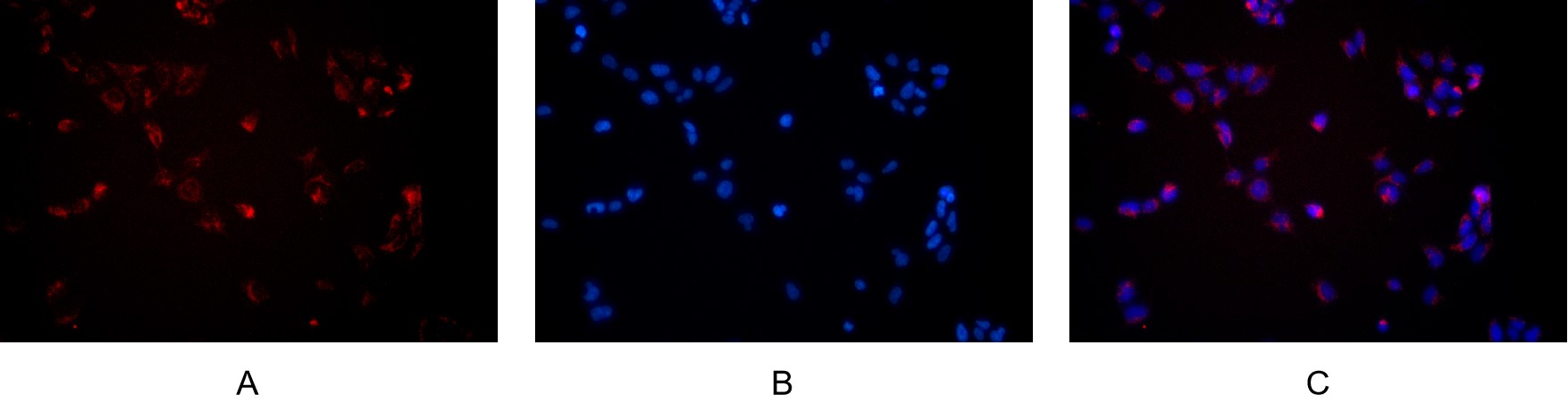
Immunofluorescence analysis of HEK293. Picture A: Mitofusin-2 antibody (red). Picture B: DAPI (blue). Picture C: Merge of A+B| Product Name | Mitofusin-2 Rabbit mAb |
|---|---|
| Antibody Type | Primary Antibodies |
| Immunogen | Recombinant Antibody expressed in animal component-free (ACF) media, purified via Protein A affinity chromatography. |
| Clonality | monoclonal |
|---|---|
| Isotype | IgG |
| Host Species | Rabbit |
| Tested Applications | ICC/IFIHCWB |
| WB:1:2000-1:10000 IHC:1:200-1:1000 ICC/IF:1:200-1:1000 |
|
| Species Reactivity | HumanMouseRat |
| Concentration | 1mg/ml |
| Purification | Protein A |
| Gene Symbol | MFN2 |
|---|---|
| Gene Synonyms | HSG MSL MARF CMT2A CPRP1 CMT2A2 HMSN6A CMT2A2A CMT2A2B |
| Gene Full Name | mitofusin 2 |
| Gene Summary | This gene encodes a mitochondrial membrane protein that participates in mitochondrial fusion and contributes to the maintenance and operation of the mitochondrial network. This protein is involved in the regulation of vascular smooth muscle cell proliferation, and it may play a role in the pathophysiology of obesity. Mutations in this gene cause Charcot-Marie-Tooth disease type 2A2, and hereditary motor and sensory neuropathy VI, which are both disorders of the peripheral nervous system. Defects in this gene have also been associated with early-onset stroke. Two transcript variants encoding the same protein have been identified. [provided by RefSeq, Jul 2008] |
| Alternative Names | MFN2 CPRP1 KIAA0214 Mitofusin-2 Transmembrane GTPase MFN2 |
| Molecular Weight(MW) | 86kD |
| Cellular Localization | Mitochondrion outer membrane |
WB
Various whole cell lysates were separated by 4-20% SDS-PAGE, and the membrane was blotted with anti-Mitofusin-2 antibody. The HRP-conjugated Goat anti-Rabbit IgG (H + L) antibody was used to detect the antibody. Lane 1: HEK293, Lane 2: SK-MEL-28, Lane 3: Mouse kidney.
IHC
Human kidney was stained with anti-Mitofusin-2 Rabbit antibody
ICC/IF
Immunofluorescence analysis of HEK293. Picture A: Mitofusin-2 antibody (red). Picture B: DAPI (blue). Picture C: Merge of A+B| Application Notes | WB:1:2000-1:10000 IHC:1:200-1:1000 ICC/IF:1:200-1:1000 |
|---|
| Form | Liquid |
|---|---|
| Storage Instructions | -15°C to -25°C/1 year(Do not lower than -25°C) |
| Storage Buffer | PBS, 50% glycerol, 0.05% Proclin 300, 0.05%BSA. |


