WB
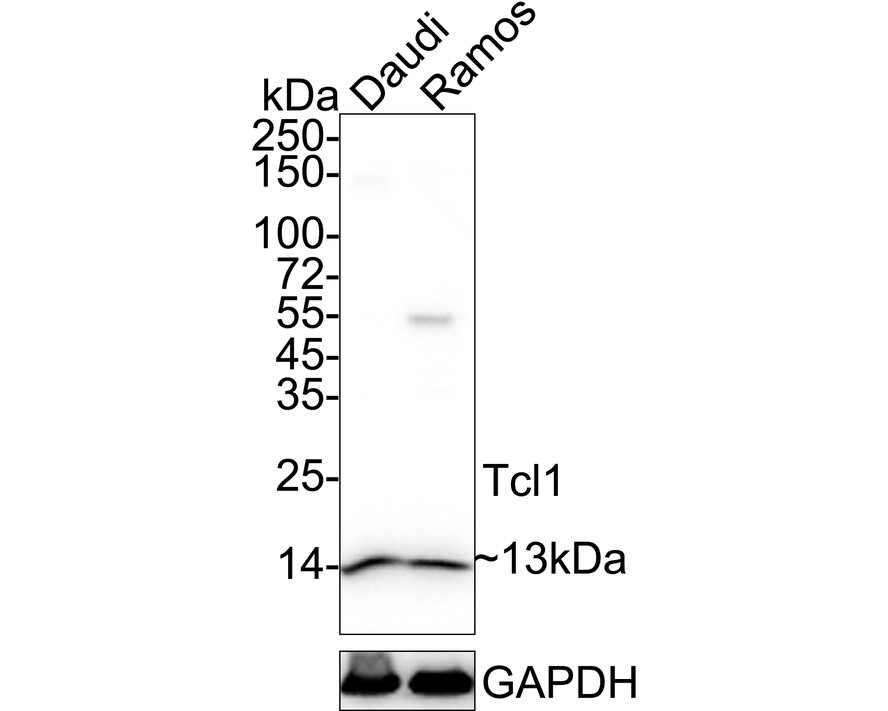
Western blot analysis of Tcl1 on different lysates with Rabbit anti-Tcl1 antibody at 1/1,000 dilution. Lane 1: Daudi cell lysate, Lane 2: Ramos cell lysate, Lysates/proteins at 20 µg/Lane. Exposure time: 10 seconds; 4-20% SDS-PAGE gel. Proteins were transferred to a PVDF membrane and blocked with 5% NFDM/TBST for 1 hour at room temperature. The primary antibody at 1/1,000 dilution was used in 5% NFDM/TBST at 4℃ overnight. Goat Anti-Rabbit IgG - HRP Secondary Antibody at 1/50,000 dilution was used for 1 hour at room temperature.IHC
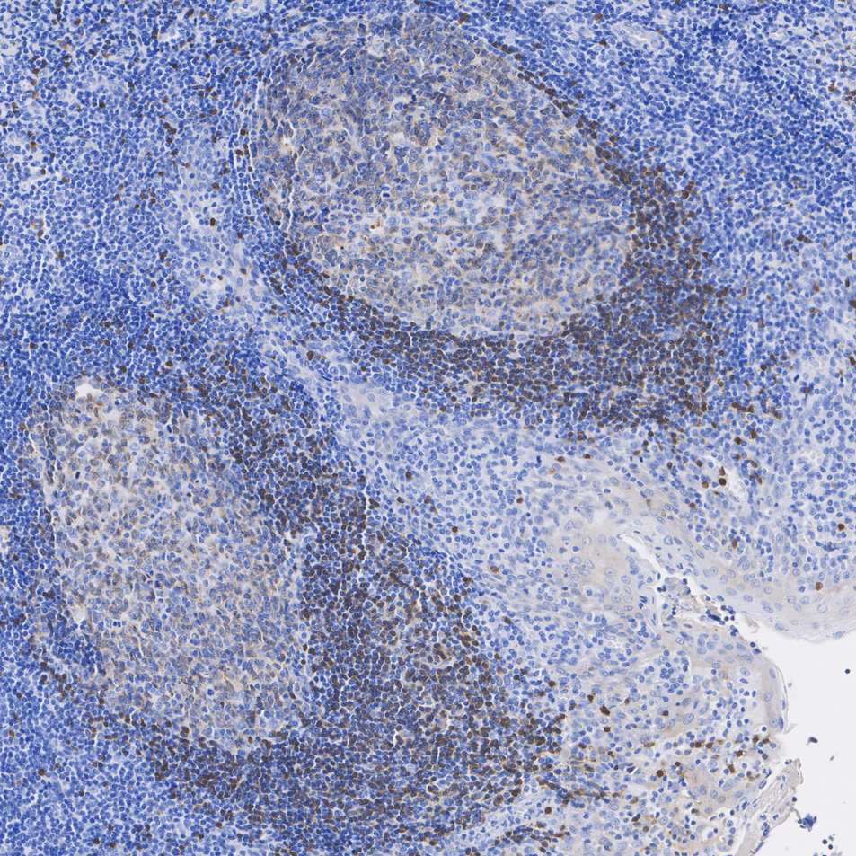
Application: Immunohistochemistry (IHC-P) Species: Human Tissue: Tonsil Sample: Paraffin-embedded section Antigen retrieval: Heat-mediated, Tris-EDTA buffer (pH 9.0) (high pressure), 2 minutes. Wash buffer: 1× TBST Endogenous peroxidase blocking: 3% H₂O₂, 10 minutes at room temperature. Blocking: 1% BSA + 10% normal goat serum, 10 minutes at room temperature. Primary antibody: 1/30,000, 1 hour at room temperature. Secondary antibody: 20 minutes at room temperature.ICC/IF
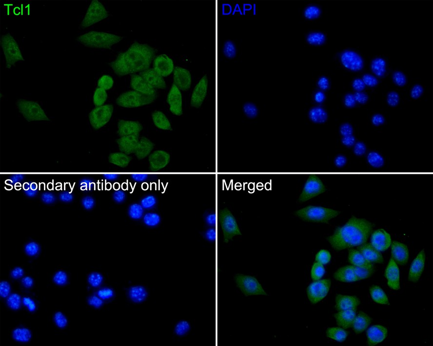
Immunocytochemistry analysis of PC-3 cells labeling Tcl1 with Rabbit anti-Tcl1 antibody at 1/50 dilution. Cells were fixed in 4% paraformaldehyde for 10 minutes at 37 ℃, permeabilized with 0.05% Triton X-100 in PBS for 20 minutes, and then blocked with 2% negative goat serum for 30 minutes at room temperature. Cells were then incubated with Rabbit anti-Tcl1 antibody at 1/50 dilution in 2% negative goat serum overnight at 4 ℃. Goat Anti-Rabbit IgG H&L was used as the secondary antibody at 1/1,000 dilution. PBS instead of the primary antibody was used as the secondary antibody only control. Nuclear DNA was labelled in blue with DAPI.FC
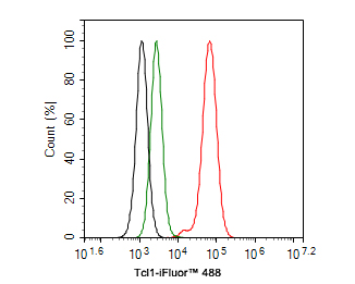
Flow cytometric analysis of Daudi cells labeling Tcl1. Cells were fixed and permeabilized. Then stained with the primary antibody (1ug/ml) (red) compared with Rabbit IgG Isotype Control (green). After incubation of the primary antibody at +4℃ for an hour, the cells were stained with a iFluor™ 488 conjugate-Goat anti-Rabbit IgG Secondary antibody at 1/1,000 dilution for 30 minutes at +4℃. Unlabelled sample was used as a control (cells without incubation with primary antibody; black).IF-F
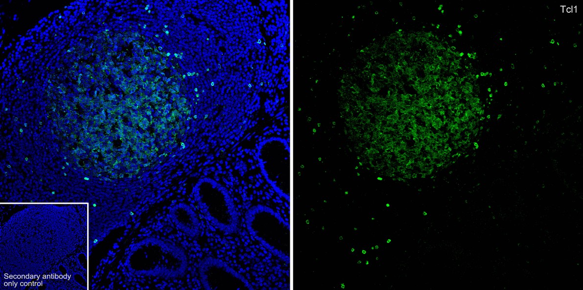
Application: IF-Tissue Species: Human Site: appendix Sample: Paraffin-embedded section Antibody concentration: 1/1,000| Product Name | Tcl1 Recombinant Rabbit Monoclonal Antibody |
|---|---|
| Antibody Type | Primary Antibodies |
| Immunogen | Recombinant protein within human Tcl1 aa 1-114. |
| Clonality | Monoclonal |
|---|---|
| Isotype | IgG |
| Host Species | Rabbit |
| Tested Applications | FCICC/IFIF-FIHCWB |
| WB:1:1000 IHC:1:10000-1:30000 ICC/IF:1:50 IF-F:1:1000 FC:1:500-1:1,000 |
|
| Species Reactivity | HumanMouseRat |
| Concentration | 1mg/ml |
| Purification | Protein A |
| Gene Symbol | TCL1A |
|---|---|
| Gene Synonyms | TCL1 |
| Gene Full Name | TCL1 family AKT coactivator A |
| Gene Summary | Overexpression of the TCL1 gene in humans has been implicated in the development of mature T cell leukemia, in which chromosomal rearrangements bring the TCL1 gene in close proximity to the T-cell antigen receptor (TCR)-alpha (MIM 186880) or TCR-beta (MIM 186930) regulatory elements (summarized by Virgilio et al., 1998 [PubMed 9520462]). In normal T cells TCL1 is expressed in CD4-/CD8- cells, but not in cells at later stages of differentiation. TCL1 functions as a coactivator of the cell survival kinase AKT (MIM 164730) (Laine et al., 2000 [PubMed 10983986]).[supplied by OMIM, Jul 2010] |
| Alternative Names | Anti TCL1A antibody Lymphoma/leukemia T-cell antibody Oncogene TCL-1 antibody Oncogene TCL1 antibody P14 TCL1 antibody P14 TCL1 protein antibody Protein p14 TCL1 antibody T cell leukemia 1 antibody T cell lymphoma 1 antibody T cell lymphoma 1A antibody Anti TCL1A antibody Lymphoma/leukemia T-cell antibody Oncogene TCL-1 antibody Oncogene TCL1 antibody P14 TCL1 antibody P14 TCL1 protein antibody Protein p14 TCL1 antibody T cell leukemia 1 antibody T cell lymphoma 1 antibody T cell lymphoma 1A antibody T-cell leukemia/lymphoma 1A antibody T-cell leukemia/lymphoma protein 1A antibody TCL 1 protein antibody TCL1 antibody TCL1 oncogene antibody TCL1 PEN antibody Tcl1a antibody TCL1A antibody antibody TCL1A_HUMAN antibody |
| Molecular Weight(MW) | 13 kDa |
| Cellular Localization | Cytoplasm, Nucleus, Microsome, Endoplasmic reticulum. |
WB
Western blot analysis of Tcl1 on different lysates with Rabbit anti-Tcl1 antibody at 1/1,000 dilution. Lane 1: Daudi cell lysate, Lane 2: Ramos cell lysate, Lysates/proteins at 20 µg/Lane. Exposure time: 10 seconds; 4-20% SDS-PAGE gel. Proteins were transferred to a PVDF membrane and blocked with 5% NFDM/TBST for 1 hour at room temperature. The primary antibody at 1/1,000 dilution was used in 5% NFDM/TBST at 4℃ overnight. Goat Anti-Rabbit IgG - HRP Secondary Antibody at 1/50,000 dilution was used for 1 hour at room temperature.
IHC
Application: Immunohistochemistry (IHC-P) Species: Human Tissue: Tonsil Sample: Paraffin-embedded section Antigen retrieval: Heat-mediated, Tris-EDTA buffer (pH 9.0) (high pressure), 2 minutes. Wash buffer: 1× TBST Endogenous peroxidase blocking: 3% H₂O₂, 10 minutes at room temperature. Blocking: 1% BSA + 10% normal goat serum, 10 minutes at room temperature. Primary antibody: 1/30,000, 1 hour at room temperature. Secondary antibody: 20 minutes at room temperature.
ICC/IF
Immunocytochemistry analysis of PC-3 cells labeling Tcl1 with Rabbit anti-Tcl1 antibody at 1/50 dilution. Cells were fixed in 4% paraformaldehyde for 10 minutes at 37 ℃, permeabilized with 0.05% Triton X-100 in PBS for 20 minutes, and then blocked with 2% negative goat serum for 30 minutes at room temperature. Cells were then incubated with Rabbit anti-Tcl1 antibody at 1/50 dilution in 2% negative goat serum overnight at 4 ℃. Goat Anti-Rabbit IgG H&L was used as the secondary antibody at 1/1,000 dilution. PBS instead of the primary antibody was used as the secondary antibody only control. Nuclear DNA was labelled in blue with DAPI.
FC
Flow cytometric analysis of Daudi cells labeling Tcl1. Cells were fixed and permeabilized. Then stained with the primary antibody (1ug/ml) (red) compared with Rabbit IgG Isotype Control (green). After incubation of the primary antibody at +4℃ for an hour, the cells were stained with a iFluor™ 488 conjugate-Goat anti-Rabbit IgG Secondary antibody at 1/1,000 dilution for 30 minutes at +4℃. Unlabelled sample was used as a control (cells without incubation with primary antibody; black).
IF-F
Application: IF-Tissue Species: Human Site: appendix Sample: Paraffin-embedded section Antibody concentration: 1/1,000| Application Notes | WB:1:1000 IHC:1:10000-1:30000 ICC/IF:1:50 IF-F:1:1000 FC:1:500-1:1,000 |
|---|
| Form | Liquid |
|---|---|
| Storage Instructions | Store at +4℃ after thawing. Aliquot store at -20℃. Avoid repeated freeze / thaw cycles. |
| Storage Buffer | 1*TBS (pH7.4), 0.05% BSA, 40% Glycerol. Preservative: 0.05% Sodium Azide. |


