WB
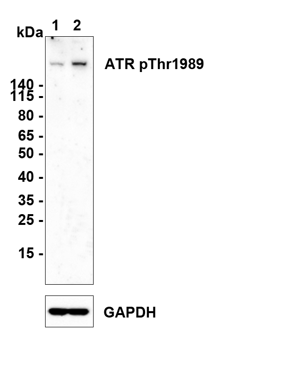
Western blot analysis of Phospho-ATR (T1989) on different lysates with Rabbit anti-Phospho-ATR (T1989) antibody at 1/1,000 dilution. Lane 1: HeLa cell lysate Lane 2: HeLa treated with Hydroxyurea cell lysate Lysates/proteins at 20 µg/Lane. Exposure time: 5 minutes; 4-20% SDS-PAGE gel. Proteins were transferred to a PVDF membrane and blocked with 5% NFDM/TBST for 1 hour at room temperature. The primary antibody at 1/1,000 dilution was used in 5% NFDM/TBST at room temperature for 2 hours. Goat Anti-Rabbit IgG - HRP Secondary Antibody at 1:300,000 dilution was used for 1 hour at room temperature.IF
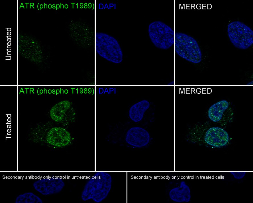
Immunocytochemistry analysis of HeLa cells treated with or without 4mM Hydroxyurea for 20 hours labeling Phospho-ATR (T1989) with Rabbit anti-Phospho-ATR (T1989) antibody at 1/200 dilution. Image shown an increased nuclear staining upon Hydroxyurea treatment. Cells were fixed in 4% paraformaldehyde for 20 minutes at 37 ℃, permeabilized with 0.1% Triton X-100 in PBS permeabilization for 5 minutes, and then blocked with 2% negative goat serum for 60 minutes at room temperature. Cells were then incubated with Rabbit anti-Phospho-ATR (T1989) antibody at 1/200 dilution in 1% BSA overnight at 4 ℃. Goat Anti-Rabbit IgG H&L was used as the secondary antibody at 1/1,000 dilution. PBS instead of the primary antibody was used as the secondary antibody only control. Nuclear DNA was labelled in blue with DAPI.| Product Name | Phospho-ATR (T1989) Recombinant Rabbit Monoclonal Antibody |
|---|---|
| Antibody Type | Primary Antibodies |
| Immunogen | Synthetic phosphopeptide corresponding to residues surrounding Thr1989 of human ATR. |
| Clonality | Monoclonal |
|---|---|
| Isotype | IgG |
| Host Species | Rabbit |
| Tested Applications | IFWB |
| WB:1:1,000 IF:1:200 |
|
| Species Reactivity | Human |
| Concentration | 1mg/ml |
| Purification | Protein A |
| Gene Symbol | ATR |
|---|---|
| Gene Synonyms | FRP1 MEC1 SCKL FCTCS SCKL1 |
| Gene Full Name | ATR checkpoint kinase |
| Gene Summary | The protein encoded by this gene is a serine/threonine kinase and DNA damage sensor, activating cell cycle checkpoint signaling upon DNA stress. The encoded protein can phosphorylate and activate several proteins involved in the inhibition of DNA replication and mitosis, and can promote DNA repair, recombination, and apoptosis. This protein is also important for fragile site stability and centrosome duplication. Defects in this gene are a cause of Seckel syndrome 1. [provided by RefSeq, Aug 2017] |
| Alternative Names | Ataxia telangiectasia and Rad3 related antibody Ataxia telangiectasia and Rad3-related protein antibody ATR antibody ATR_HUMAN antibody FCTCS antibody FRAP Related Protein 1 antibody FRAP-related protein 1 antibody FRP1 antibody MEC1 antibody MEC1 mitosis entry checkpoint 1 homolog antibody Ataxia telangiectasia and Rad3 related antibody Ataxia telangiectasia and Rad3-related protein antibody ATR antibody ATR_HUMAN antibody FCTCS antibody FRAP Related Protein 1 antibody FRAP-related protein 1 antibody FRP1 antibody MEC1 antibody MEC1 mitosis entry checkpoint 1 homolog antibody Protein kinase ATR antibody RAC3 antibody Rad3 related protein antibody SCKL antibody SCKL1 antibody Serine/threonine protein kinase ATR antibody Serine/threonine-protein kinase ATR antibody |
| Molecular Weight(MW) | 301 kDa |
| Cellular Localization | Chromosome,Nucleus |
WB
Western blot analysis of Phospho-ATR (T1989) on different lysates with Rabbit anti-Phospho-ATR (T1989) antibody at 1/1,000 dilution. Lane 1: HeLa cell lysate Lane 2: HeLa treated with Hydroxyurea cell lysate Lysates/proteins at 20 µg/Lane. Exposure time: 5 minutes; 4-20% SDS-PAGE gel. Proteins were transferred to a PVDF membrane and blocked with 5% NFDM/TBST for 1 hour at room temperature. The primary antibody at 1/1,000 dilution was used in 5% NFDM/TBST at room temperature for 2 hours. Goat Anti-Rabbit IgG - HRP Secondary Antibody at 1:300,000 dilution was used for 1 hour at room temperature.
IF
Immunocytochemistry analysis of HeLa cells treated with or without 4mM Hydroxyurea for 20 hours labeling Phospho-ATR (T1989) with Rabbit anti-Phospho-ATR (T1989) antibody at 1/200 dilution. Image shown an increased nuclear staining upon Hydroxyurea treatment. Cells were fixed in 4% paraformaldehyde for 20 minutes at 37 ℃, permeabilized with 0.1% Triton X-100 in PBS permeabilization for 5 minutes, and then blocked with 2% negative goat serum for 60 minutes at room temperature. Cells were then incubated with Rabbit anti-Phospho-ATR (T1989) antibody at 1/200 dilution in 1% BSA overnight at 4 ℃. Goat Anti-Rabbit IgG H&L was used as the secondary antibody at 1/1,000 dilution. PBS instead of the primary antibody was used as the secondary antibody only control. Nuclear DNA was labelled in blue with DAPI.| Application Notes | WB:1:1,000 IF:1:200 |
|---|
| Form | Liquid |
|---|---|
| Storage Instructions | Shipped at 4℃. Store at +4℃ short term (1-2 weeks). Store at -20℃ long term. |
| Storage Buffer | PBS (pH7.4), 0.1% BSA, 40% Glycerol. Preservative: 0.05% Sodium Azide. |


