WB
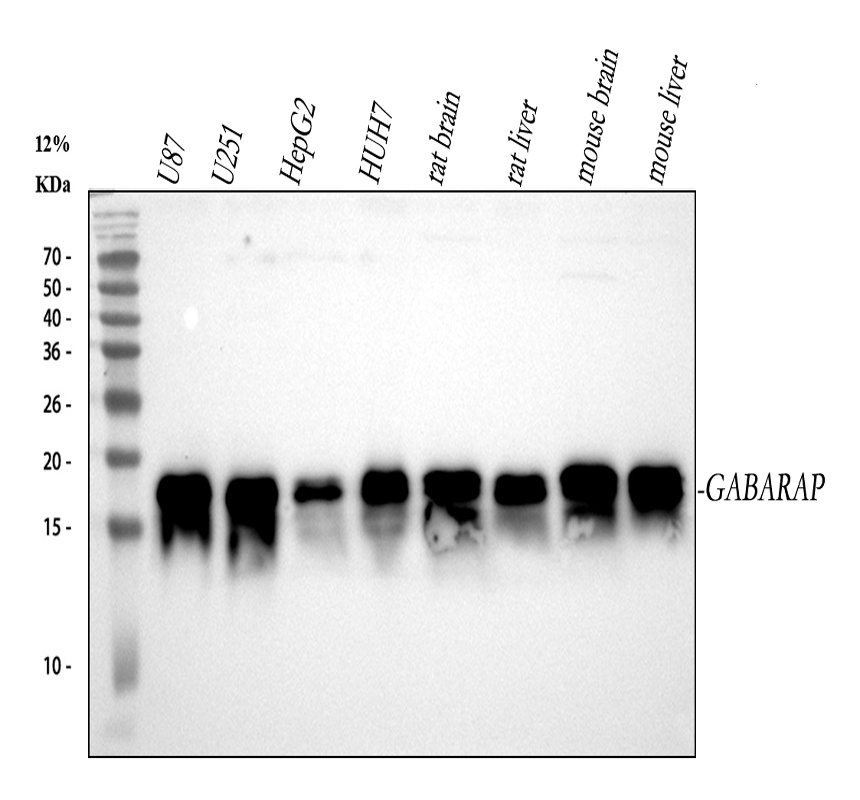
Western blot analysis of anti-GABARAP antibody. The sample well of each lane was loaded with 30 ug of sample under reducing conditions. Lane 1: human U87 whole cell lysates, Lane 2: human U251 whole cell lysates, Lane 3: human HepG2 whole cell lysates, Lane 4: human HUH7 whole cell lysates, Lane 5: rat brain tissue lysates, Lane 6: rat liver tissue lysates, Lane 7: mouse brain tissue lysates, Lane 8: mouse liver tissue lysates. After electrophoresis, proteins were transferred to a membrane. Then the membrane was incubated with rabbit anti-GABARAP antigen affinity purified monoclonal antibody at a dilution of 1:1000 and probed with a goat anti-rabbit IgG-HRP secondary antibody. The signal is developed using ECL Plus Western Blotting Substrate. A specific band was detected for GABARAP at approximately 18 kDa. The expected band size for GABARAP is at 14 kDa.| Product Name | Anti-GABARAP Antibody |
|---|---|
| Antibody Type | Primary Antibodies |
| Immunogen | A synthesized peptide derived from human GABARAP GABARAP is cleaved at its carboxyl terminus, which leads to conjugation by either of the phospholipids phosphatidylethanolamine or phosphatidylserine. This processing converts GABARAP from a type I to a type II membrane bound form involved in autophagosome biogenesis. Processing of GABARAP involves cleavage by Atg4 family members followed by conjugation by the E1 and E2 like enzymes Atg7 and Atg3. |
| Clonality | monoclonal |
|---|---|
| Isotype | IgG |
| Host Species | Rabbit |
| Tested Applications | FCICC/IFIHCWB |
| WB:1:1000-10000 IHC:1:50-200 ICC/IF:1:50-200 FC:1:50 |
|
| Species Reactivity | HumanMouseRabbit |
| Concentration | 0.5mg/ml |
| Purification | Affinity purified |
| Gene Symbol | GABARAP |
|---|---|
| Gene Synonyms | MM46 ATG8A GABARAP-a |
| Gene Full Name | GABA type A receptor-associated protein |
| Gene Summary | Gamma-aminobutyric acid A receptors [GABA(A) receptors] are ligand-gated chloride channels that mediate inhibitory neurotransmission. This gene encodes GABA(A) receptor-associated protein, which is highly positively charged in its N-terminus and shares sequence similarity with light chain-3 of microtubule-associated proteins 1A and 1B. This protein clusters neurotransmitter receptors by mediating interaction with the cytoskeleton. [provided by RefSeq, Jul 2008] |
| Alternative Names | FLC3B GABARAP MM46 |
| Molecular Weight(MW) | 14 kDa(Observed band size:18 kDa) |
| Cellular Localization | Cytoplasm,Cytoplasmic vesicle,Cytoskeleton,Golgi apparatus,Membrane,Microtubule |
WB
Western blot analysis of anti-GABARAP antibody. The sample well of each lane was loaded with 30 ug of sample under reducing conditions. Lane 1: human U87 whole cell lysates, Lane 2: human U251 whole cell lysates, Lane 3: human HepG2 whole cell lysates, Lane 4: human HUH7 whole cell lysates, Lane 5: rat brain tissue lysates, Lane 6: rat liver tissue lysates, Lane 7: mouse brain tissue lysates, Lane 8: mouse liver tissue lysates. After electrophoresis, proteins were transferred to a membrane. Then the membrane was incubated with rabbit anti-GABARAP antigen affinity purified monoclonal antibody at a dilution of 1:1000 and probed with a goat anti-rabbit IgG-HRP secondary antibody. The signal is developed using ECL Plus Western Blotting Substrate. A specific band was detected for GABARAP at approximately 18 kDa. The expected band size for GABARAP is at 14 kDa.| Application Notes | WB:1:1000-10000 IHC:1:50-200 ICC/IF:1:50-200 FC:1:50 |
|---|
| Form | Liquid |
|---|---|
| Storage Instructions | 12 months from date of receipt,-20℃ as supplied. |
| Storage Buffer | 500 ug/ml; Rabbit IgG in phosphate buffered saline, pH 7.4, 150mM NaCl, 0.02% sodium azide, 0.4-0.5 mg/ml BSA and 50% glycerol. |


