WB
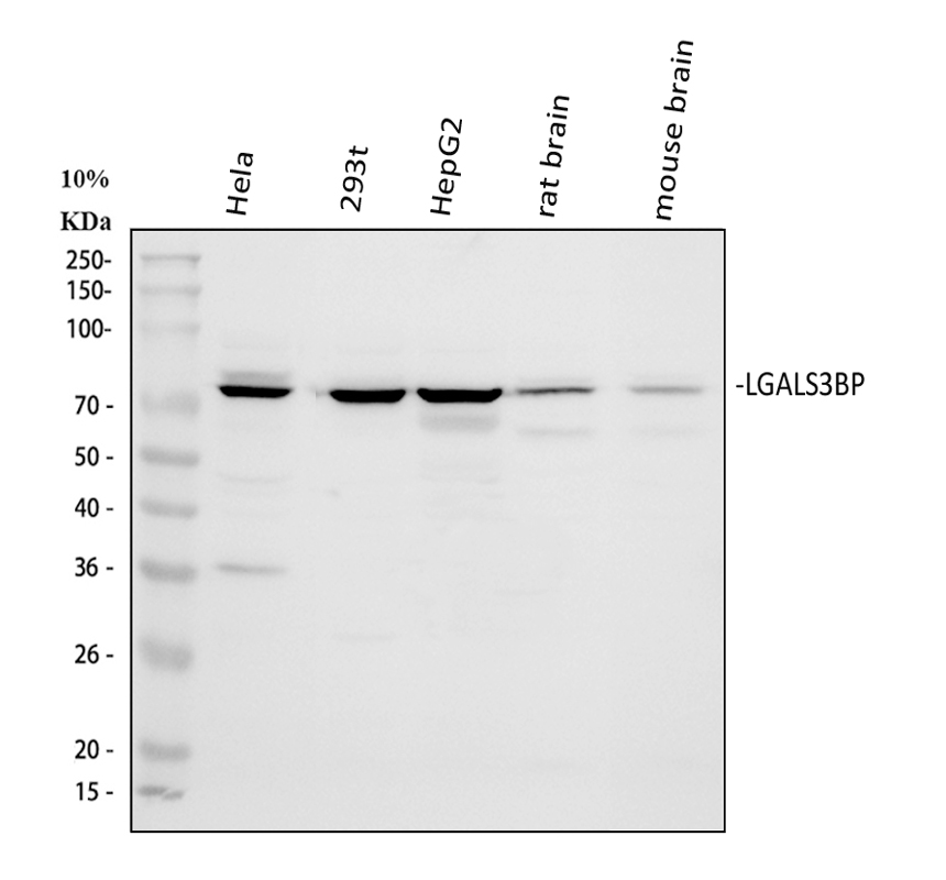
Western blot analysis of LGALS3BP using anti-LGALS3BP antibody. The sample well of each lane was loaded with 30 ug of sample under reducing conditions. Lane 1: human Hela whole cell lysates, Lane 2: human 293T whole cell lysates, Lane 3: human HepG2 whole cell lysates, Lane 4: rat brain tissue lysates, Lane 5: mouse brain tissue lysates. After electrophoresis, proteins were transferred to a membrane. Then the membrane was incubated with rabbit anti-LGALS3BP antigen affinity purified polyclonal antibody at a dilution of 1:1000 and probed with a goat anti-rabbit IgG-HRP secondary antibody. The signal is developed using ECL Plus Western Blotting Substrate. A specific band was detected for LGALS3BP at approximately 75 kDa. The expected band size for LGALS3BP is at 65 kDa.IHC
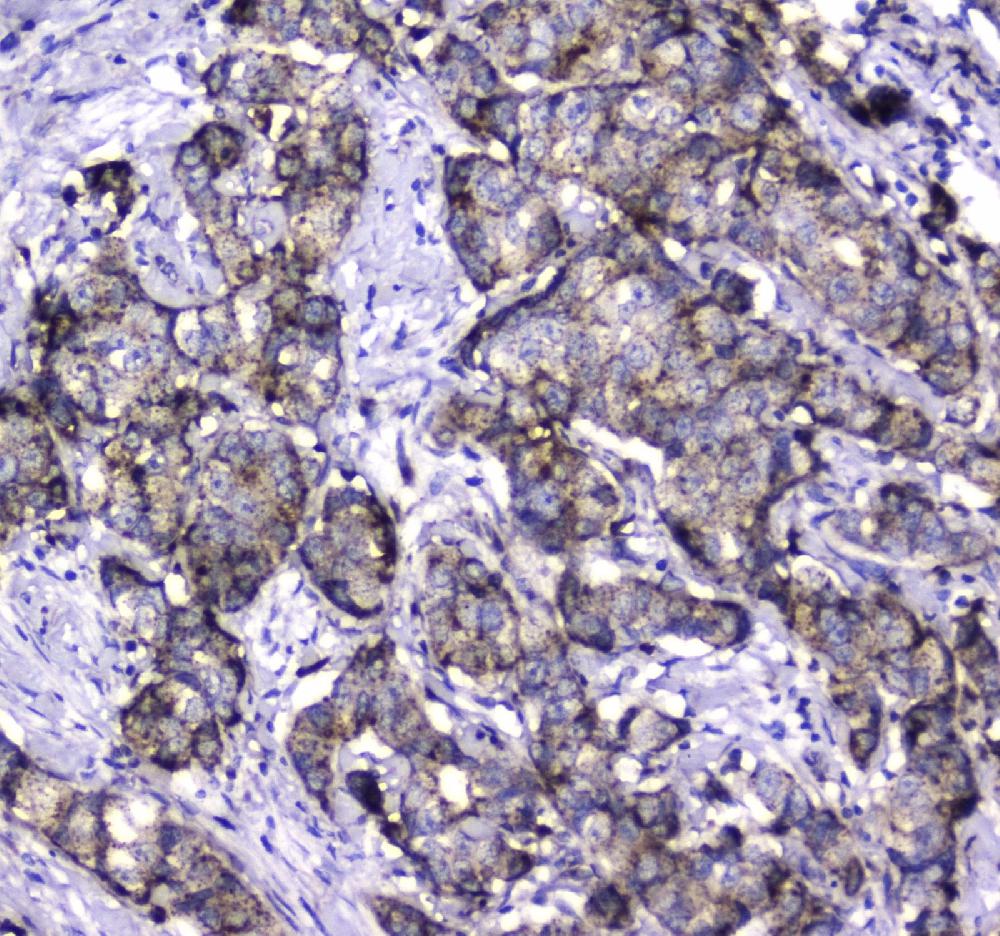
IHC analysis of LGALS3BP using anti-LGALS3BP antibody. LGALS3BP was detected in a paraffin-embedded section of human mammary cancer tissue. Biotinylated goat anti-rabbit IgG was used as secondary antibody. The tissue section was incubated with rabbit anti-LGALS3BP Antibody at a dilution of 1:200 and developed using Strepavidin-Biotin-Complex (SABC) with DAB as the chromogen.IHC
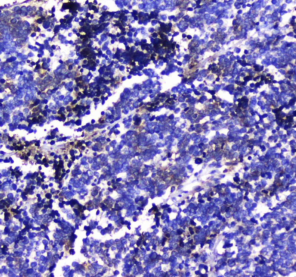
IHC analysis of LGALS3BP using anti-LGALS3BP antibody. LGALS3BP was detected in a paraffin-embedded section of human lung cancer tissue. Biotinylated goat anti-rabbit IgG was used as secondary antibody. The tissue section was incubated with rabbit anti-LGALS3BP Antibody at a dilution of 1:200 and developed using Strepavidin-Biotin-Complex (SABC) with DAB as the chromogen.| Product Name | Anti-LGALS3BP Antibody |
|---|---|
| Antibody Type | Primary Antibodies |
| Immunogen | A synthetic peptide corresponding to a sequence in the middle region of human LGALS3BP, which shares 82.1% and 71.8% amino acid (aa) sequence identity with mouse and rat LGALS3BP, respectively. |
| Clonality | Polyclonal |
|---|---|
| Isotype | IgG |
| Host Species | Rabbit |
| Tested Applications | IHCWB |
| WB:1:500-1:2000 IHC:1:50-400 |
|
| Species Reactivity | HumanMouseRat |
| Concentration | 0.5mg/ml |
| Purification | Affinity purified |
| Gene Symbol | LGALS3BP |
|---|---|
| Gene Synonyms | 90K M2BP gp90 CyCAP BTBD17B MAC-2-BP TANGO10B |
| Gene Full Name | galectin 3 binding protein |
| Gene Summary | The galectins are a family of beta-galactoside-binding proteins implicated in modulating cell-cell and cell-matrix interactions. LGALS3BP has been found elevated in the serum of patients with cancer and in those infected by the human immunodeficiency virus (HIV). It appears to be implicated in immune response associated with natural killer (NK) and lymphokine-activated killer (LAK) cell cytotoxicity. Using fluorescence in situ hybridization the full length 90K cDNA has been localized to chromosome 17q25. The native protein binds specifically to a human macrophage-associated lectin known as Mac-2 and also binds galectin 1. [provided by RefSeq, Jul 2008] |
| Alternative Names | 90K BTBD17B Galectin 3 binding protein LGALS3BP M2BP Mac 2 binding protein MAC 2 BP MAC2BP Tumor associated antigen 90K |
| Molecular Weight(MW) | 65 kDa |
| Cellular Localization | Extracellular matrix,Secreted |
WB
Western blot analysis of LGALS3BP using anti-LGALS3BP antibody. The sample well of each lane was loaded with 30 ug of sample under reducing conditions. Lane 1: human Hela whole cell lysates, Lane 2: human 293T whole cell lysates, Lane 3: human HepG2 whole cell lysates, Lane 4: rat brain tissue lysates, Lane 5: mouse brain tissue lysates. After electrophoresis, proteins were transferred to a membrane. Then the membrane was incubated with rabbit anti-LGALS3BP antigen affinity purified polyclonal antibody at a dilution of 1:1000 and probed with a goat anti-rabbit IgG-HRP secondary antibody. The signal is developed using ECL Plus Western Blotting Substrate. A specific band was detected for LGALS3BP at approximately 75 kDa. The expected band size for LGALS3BP is at 65 kDa.
IHC
IHC analysis of LGALS3BP using anti-LGALS3BP antibody. LGALS3BP was detected in a paraffin-embedded section of human mammary cancer tissue. Biotinylated goat anti-rabbit IgG was used as secondary antibody. The tissue section was incubated with rabbit anti-LGALS3BP Antibody at a dilution of 1:200 and developed using Strepavidin-Biotin-Complex (SABC) with DAB as the chromogen.
IHC
IHC analysis of LGALS3BP using anti-LGALS3BP antibody. LGALS3BP was detected in a paraffin-embedded section of human lung cancer tissue. Biotinylated goat anti-rabbit IgG was used as secondary antibody. The tissue section was incubated with rabbit anti-LGALS3BP Antibody at a dilution of 1:200 and developed using Strepavidin-Biotin-Complex (SABC) with DAB as the chromogen.| Application Notes | WB:1:500-1:2000 IHC:1:50-400 |
|---|
| Form | Liquid |
|---|---|
| Storage Instructions | 12 months from date of receipt, -20℃ as supplied. 6 months 2 to 8℃ after reconstitution. Avoid repeated freezing and thawing. |
| Storage Buffer | 500ug/ml antibody with PBS, 0.02% NaN3, 1 mg/ml BSA and 50% glycerol. |


