WB
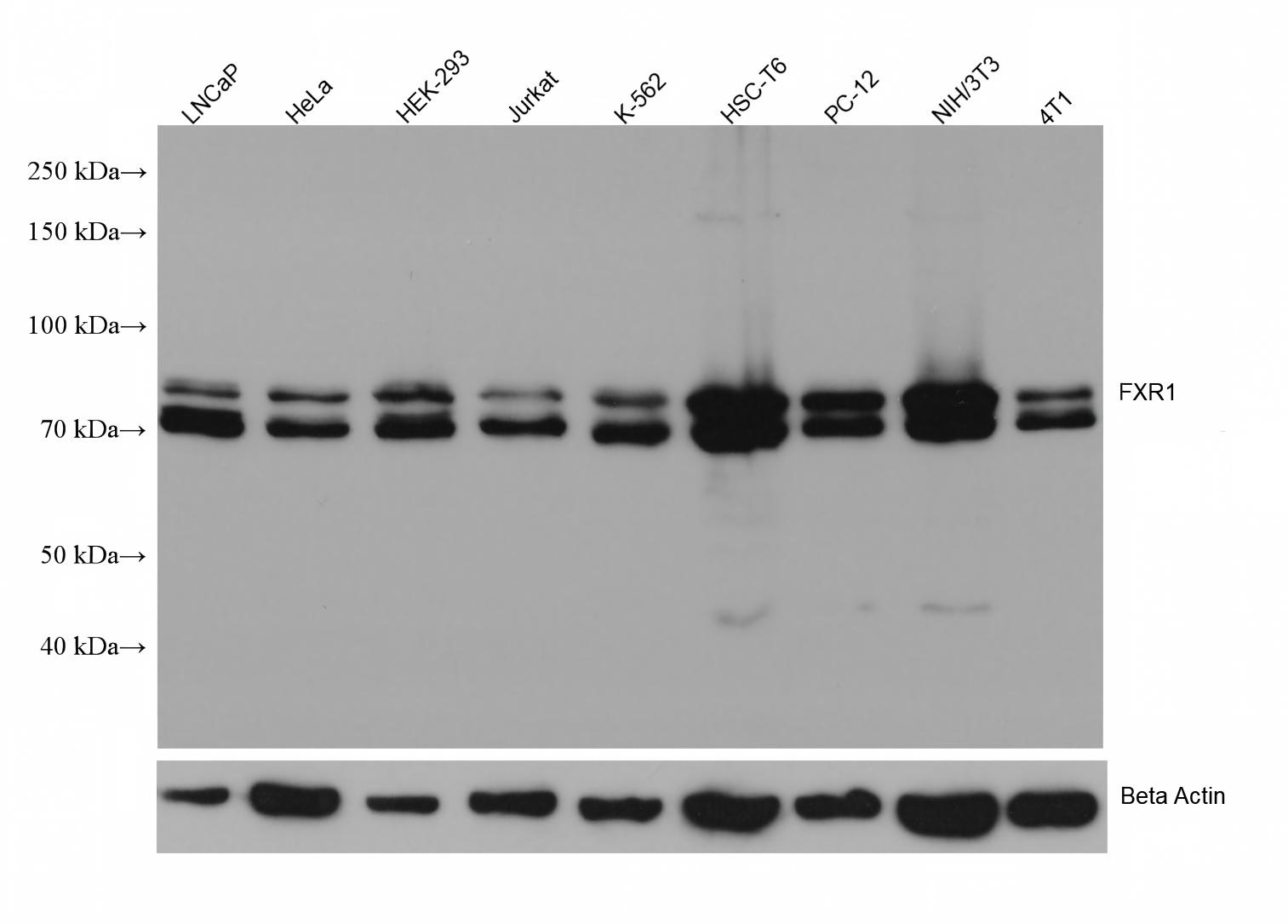
Various lysates were subjected to SDS PAGE followed by western blot with FXR1 antibody at dilution of 1:10000 incubated at room temperature for 1.5 hours. The membrane was stripped and reblotted with HRP-conjugated Beta Actin Monoclonal antibody as loading control.IHC
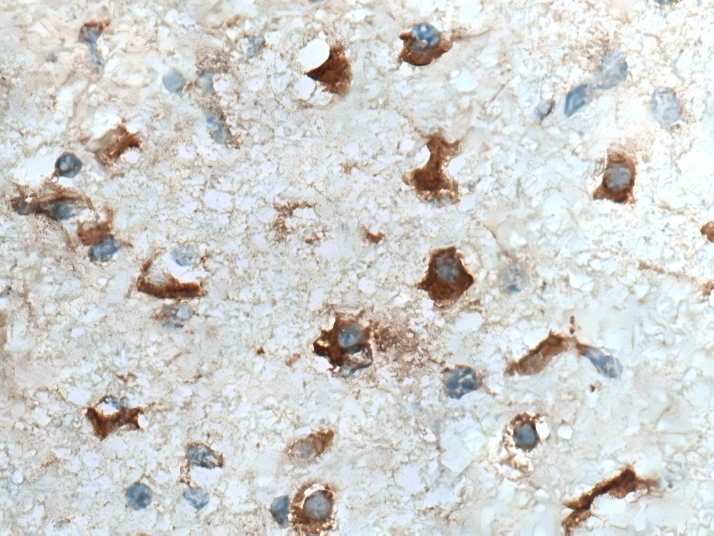
Immunohistochemical analysis of paraffin-embedded human gliomas tissue slide using FXR1 antibody at dilution of 1:1000 (under 40x lens). Heat mediated antigen retrieval with Tris-EDTA buffer (pH 9.0).IHC
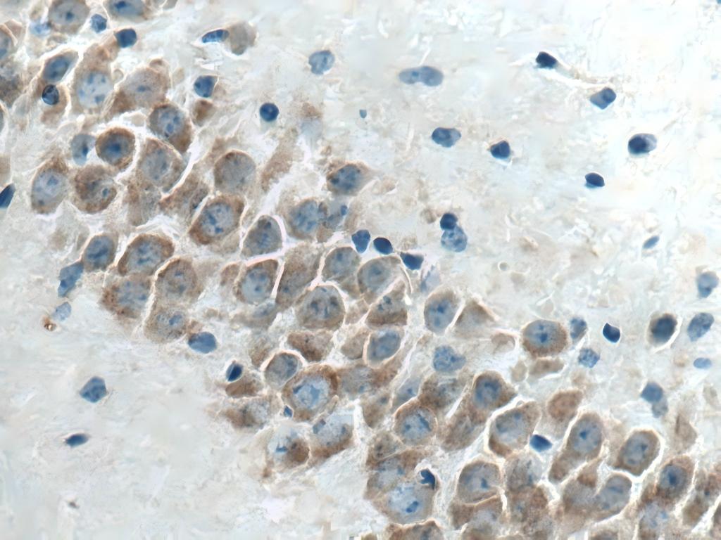
Immunohistochemical analysis of paraffin-embedded mouse brain tissue slide using FXR1 antibody at dilution of 1:1000 (under 40x lens). Heat mediated antigen retrieval with Tris-EDTA buffer (pH 9.0).IP
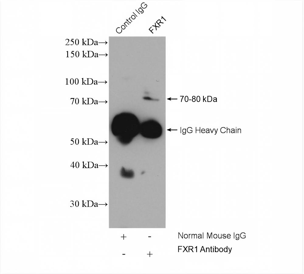
IP result of anti-FXR1 (IP:5ug; Detection:1:1000) with K-562 cells lysate 3680 ug.ICC/IF
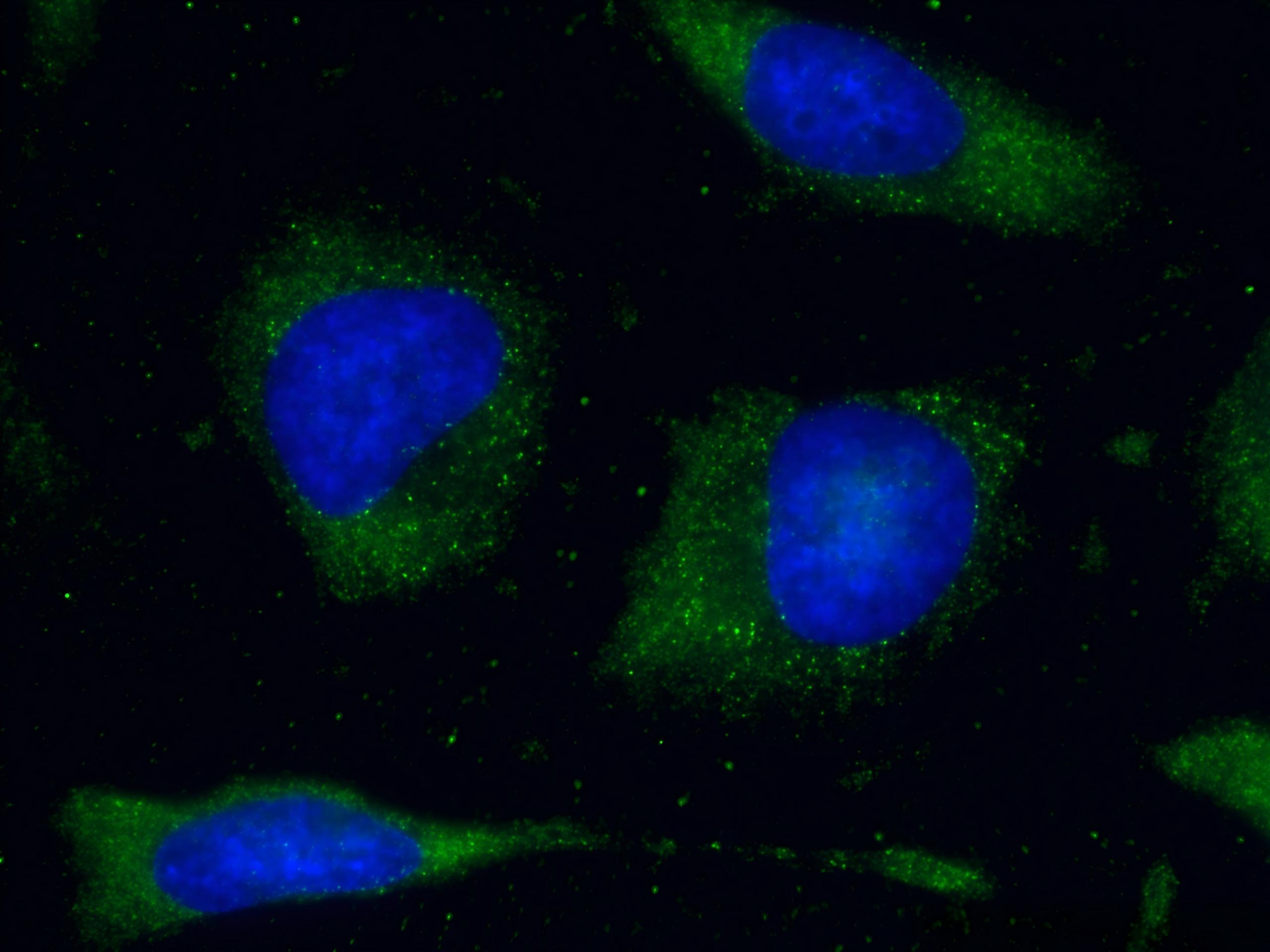
Immunofluorescent analysis of (-20°C Methanol) fixed HeLa cells using FXR1 antibody at dilution of 1:400 and CoraLite®488-Conjugated AffiniPure Goat Anti-Mouse IgG(H+L).| Product Name | FXR1 Monoclonal antibody |
|---|---|
| Antibody Type | Primary Antibodies |
| Immunogen | Recombinant human FXR1 protein |
| Clonality | Monoclonal |
|---|---|
| Isotype | IgG1 |
| Host Species | Mouse |
| Tested Applications | ICC/IFIHCIPWB |
| WB:1:5000-1:50000 IHC :1:500-1:2000 IP :0.5-4.0 ug for 1.0-3.0 mg of total protein lysate ICC/IF :1:200-1:800 |
|
| Species Reactivity | HumanMouseRat |
| Concentration | 1mg/ml |
| Purification | Protein G |
| Gene Symbol | FXR1 |
|---|---|
| Gene Synonyms | FXR1P CMYO9A CMYO9B CMYP9A CMYP9B MYOPMIL MYORIBF |
| Gene Full Name | FMR1 autosomal homolog 1 |
| Gene Summary | The protein encoded by this gene is an RNA binding protein that interacts with the functionally-similar proteins FMR1 and FXR2. These proteins shuttle between the nucleus and cytoplasm and associate with polyribosomes, predominantly with the 60S ribosomal subunit. Three transcript variants encoding different isoforms have been found for this gene. [provided by RefSeq, Jul 2008] |
| Alternative Names | 1E12E8 FMR1 autosomal homolog 1 hFXR1p RNA-binding protein FXR1 |
| Molecular Weight(MW) | 70 kDa (Observed band size: 70-80kDa) |
| Cellular Localization | Cell projection、Cytoplasm、Nucleus、Synapse |
WB
Various lysates were subjected to SDS PAGE followed by western blot with FXR1 antibody at dilution of 1:10000 incubated at room temperature for 1.5 hours. The membrane was stripped and reblotted with HRP-conjugated Beta Actin Monoclonal antibody as loading control.
IHC
Immunohistochemical analysis of paraffin-embedded human gliomas tissue slide using FXR1 antibody at dilution of 1:1000 (under 40x lens). Heat mediated antigen retrieval with Tris-EDTA buffer (pH 9.0).
IHC
Immunohistochemical analysis of paraffin-embedded mouse brain tissue slide using FXR1 antibody at dilution of 1:1000 (under 40x lens). Heat mediated antigen retrieval with Tris-EDTA buffer (pH 9.0).
IP
IP result of anti-FXR1 (IP:5ug; Detection:1:1000) with K-562 cells lysate 3680 ug.
ICC/IF
Immunofluorescent analysis of (-20°C Methanol) fixed HeLa cells using FXR1 antibody at dilution of 1:400 and CoraLite®488-Conjugated AffiniPure Goat Anti-Mouse IgG(H+L).| Application Notes | WB:1:5000-1:50000 IHC :1:500-1:2000 IP :0.5-4.0 ug for 1.0-3.0 mg of total protein lysate ICC/IF :1:200-1:800 |
|---|
| Form | Liquid |
|---|---|
| Storage Instructions | Store at -20°C. Stable for one year after shipment. |
| Storage Buffer | PBS with 0.02% sodium azide and 50% glycerol pH 7.3. |


