WB
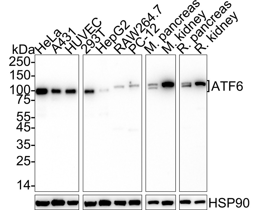
Western blot analysis of ATF6 on different lysates with Rabbit anti-ATF6 antibody at 1/1000 dilution. Lane 1: HeLa cell lysate, Lane 2: A431 cell lysate, Lane 3: HUVEC cell lysate, Lane 4: 293T cell lysate, Lane 5: HepG2 cell lysate, Lane 6: RAW264.7 cell lysate, Lane 7: PC-12 cell lysate, Lane 8: Mouse pancreas tissue lysate, Lane 9: Mouse kidney tissue lysate, Lane 10: Rat pancreas tissue lysate, Lane 11: Rat kidney tissue lysate, Lysates/proteins at 20 µg/Lane. Exposure time: 2 minutes; 4-20% SDS-PAGE gel. Proteins were transferred to a PVDF membrane and blocked with 5% NFDM/TBST for 1 hour at room temperature. The primary antibody at 1/1000 dilution was used in 5% NFDM/TBST at 4℃ overnight. Goat Anti-Rabbit IgG - HRP Secondary Antibody at 1/50000 dilution was used for 1 hour at room temperature.IHC
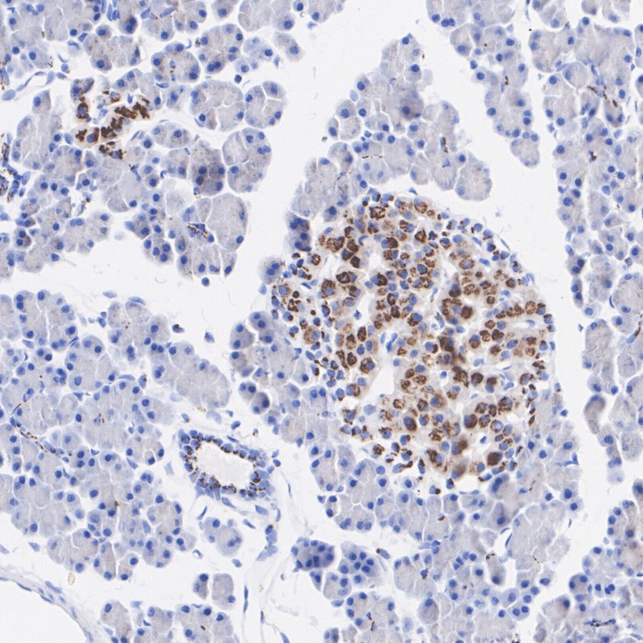
Immunohistochemical analysis of paraffin-embedded rat pancreas tissue with Rabbit anti-ATF6 antibody at 1/1000 dilution. The section was pre-treated using heat mediated antigen retrieval with Tris-EDTA buffer (pH 9.0) for 20 minutes. The tissues were blocked in 1% BSA for 20 minutes at room temperature, washed with ddH2O and PBS, and then probed with the primary antibody at 1/1000 dilution for 1 hour at room temperature. The detection was performed using an HRP conjugated compact polymer system. DAB was used as the chromogen. Tissues were counterstained with hematoxylin and mounted with DPX.ChIP
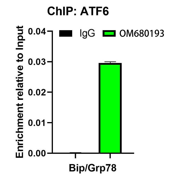
Chromatin immunoprecipitations were performed with cross-linked chromatin from HeLa cells treated with 1μM Thapsigargin for 1 hour with ATF6 or Normal Rabbit IgG according to the ChIP protocol. The enriched DNA was quantified by real-time PCR using indicated primers. The amount of immunoprecipitated DNA in each sample is represented as signal relative to the total amount of input chromatin, which is equivalent to one.| Product Name | ATF6 Recombinant Rabbit Monoclonal Antibody |
|---|---|
| Antibody Type | Primary Antibodies |
| Product description |
|
| Immunogen | Recombinant protein within human ATF6 aa 1-350. |
| Clonality | monoclonal |
|---|---|
| Isotype | IgG |
| Host Species | Rabbit |
| Tested Applications | ChIPIHCWB |
| WB:1:1000 IHC:1:200-1:1000 ChIP:Use 0.5~2 μg for 25 μg of chromatin. |
|
| Species Reactivity | HumanMouseRat |
| Concentration | 1mg/ml |
| Purification | Protein A |
| Gene Symbol | ATF6 |
|---|---|
| Gene Synonyms | ACHM7 ATF6A ATP6alpha |
| Gene Full Name | activating transcription factor 6 |
| Gene Summary | This gene encodes a transcription factor that activates target genes for the unfolded protein response (UPR) during endoplasmic reticulum (ER) stress. Although it is a transcription factor, this protein is unusual in that it is synthesized as a transmembrane protein that is embedded in the ER. It functions as an ER stress sensor/transducer, and following ER stress-induced proteolysis, it functions as a nuclear transcription factor via a cis-acting ER stress response element (ERSE) that is present in the promoters of genes encoding ER chaperones. This protein has been identified as a survival factor for quiescent but not proliferative squamous carcinoma cells. There have been conflicting reports about the association of polymorphisms in this gene with diabetes in different populations, but another polymorphism has been associated with increased plasma cholesterol levels. This gene is also thought to be a potential therapeutic target for cystic fibrosis. [provided by RefSeq, Aug 2011] |
| Alternative Names | Activating transcription factor 6 alpha antibody Activating transcription factor 6 antibody ATF 6 antibody ATF6 alpha antibody ATF6 antibody ATF6-alpha antibody ATF6A antibody ATF6A_HUMAN antibody cAMP dependent transcription factor ATF 6 alpha antibody cAMP-dependent transcription factor ATF-6 alpha antibody Activating transcription factor 6 alpha antibody Activating transcription factor 6 antibody ATF 6 antibody ATF6 alpha antibody ATF6 antibody ATF6-alpha antibody ATF6A antibody ATF6A_HUMAN antibody cAMP dependent transcription factor ATF 6 alpha antibody cAMP-dependent transcription factor ATF-6 alpha antibody Cyclic AMP dependent transcription factor ATF 6 alpha antibody DKFZp686P2194 antibody ESTM49 antibody FLJ21663 antibody Processed cyclic AMP dependent transcription factor ATF 6 alpha antibody Processed cyclic AMP-dependent transcription factor ATF-6 alpha antibody |
| Molecular Weight(MW) | 75kDa(Observed band size:100-110kDa) |
| Cellular Localization | Endoplasmic reticulum membrane, Golgi apparatus membrane. |
WB
Western blot analysis of ATF6 on different lysates with Rabbit anti-ATF6 antibody at 1/1000 dilution. Lane 1: HeLa cell lysate, Lane 2: A431 cell lysate, Lane 3: HUVEC cell lysate, Lane 4: 293T cell lysate, Lane 5: HepG2 cell lysate, Lane 6: RAW264.7 cell lysate, Lane 7: PC-12 cell lysate, Lane 8: Mouse pancreas tissue lysate, Lane 9: Mouse kidney tissue lysate, Lane 10: Rat pancreas tissue lysate, Lane 11: Rat kidney tissue lysate, Lysates/proteins at 20 µg/Lane. Exposure time: 2 minutes; 4-20% SDS-PAGE gel. Proteins were transferred to a PVDF membrane and blocked with 5% NFDM/TBST for 1 hour at room temperature. The primary antibody at 1/1000 dilution was used in 5% NFDM/TBST at 4℃ overnight. Goat Anti-Rabbit IgG - HRP Secondary Antibody at 1/50000 dilution was used for 1 hour at room temperature.
IHC
Immunohistochemical analysis of paraffin-embedded rat pancreas tissue with Rabbit anti-ATF6 antibody at 1/1000 dilution. The section was pre-treated using heat mediated antigen retrieval with Tris-EDTA buffer (pH 9.0) for 20 minutes. The tissues were blocked in 1% BSA for 20 minutes at room temperature, washed with ddH2O and PBS, and then probed with the primary antibody at 1/1000 dilution for 1 hour at room temperature. The detection was performed using an HRP conjugated compact polymer system. DAB was used as the chromogen. Tissues were counterstained with hematoxylin and mounted with DPX.
ChIP
Chromatin immunoprecipitations were performed with cross-linked chromatin from HeLa cells treated with 1μM Thapsigargin for 1 hour with ATF6 or Normal Rabbit IgG according to the ChIP protocol. The enriched DNA was quantified by real-time PCR using indicated primers. The amount of immunoprecipitated DNA in each sample is represented as signal relative to the total amount of input chromatin, which is equivalent to one.| Application Notes | WB:1:1000 IHC:1:200-1:1000 ChIP:Use 0.5~2 μg for 25 μg of chromatin. |
|---|
| Form | Liquid |
|---|---|
| Storage Instructions | Shipped at 4℃. Store at +4℃ short term (1-2 weeks). It is recommended to aliquot into single-use upon delivery. Store at -20℃ long term. |
| Storage Buffer | PBS (pH7.4), 0.1% BSA, 40% Glycerol. Preservative: 0.05% Sodium Azide. |


