
WB
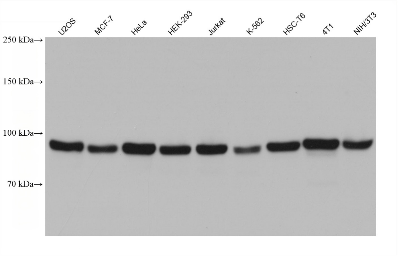
Various lysates were subjected to SDS PAGE followed by western blot with ATF6 antibody at dilution of 1:10000 incubated at room temperature for 1.5 hours.WB
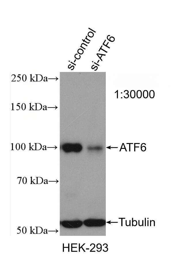
WB result of ATF6 antibody (1:30000; incubated at room temperature for 1.5 hours) with sh-Control and sh-ATF6 transfected HEK-293 cells.WB
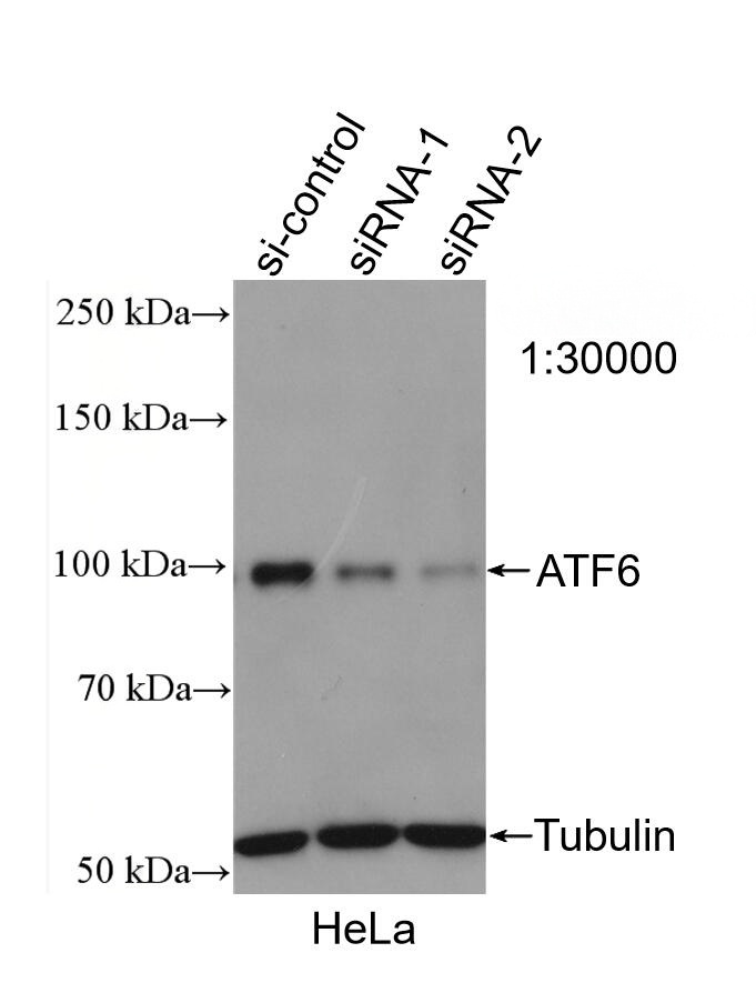
WB result of ATF6 antibody (1:30000; incubated at room temperature for 1.5 hours) with sh-Control and sh-ATF6 transfected HeLa cells.IHC
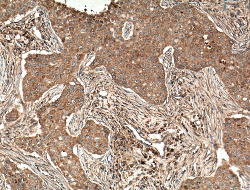
Immunohistochemical analysis of paraffin-embedded human breast cancer tissue slide using ATF6 antibody at dilution of 1:500 (under 10x lens. Heat mediated antigen retrieval with Tris-EDTA buffer (pH 9.0).IHC
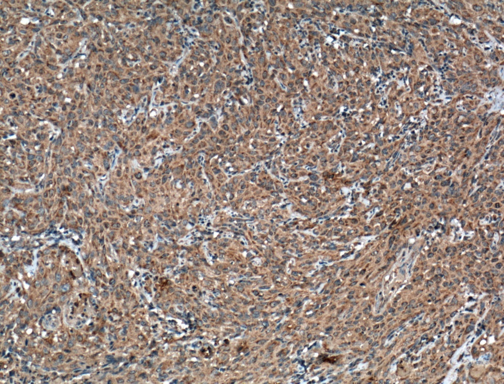
Immunohistochemical analysis of paraffin-embedded human cervical cancer tissue slide using ATF6 antibody at dilution of 1:500 (under 10x lens. Heat mediated antigen retrieval with Tris-EDTA buffer (pH 9.0).IHC
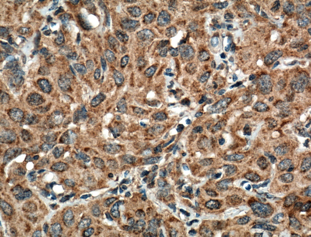
Immunohistochemical analysis of paraffin-embedded human cervical cancer tissue slide using ATF6 antibody at dilution of 1:500 (under 40x lens. Heat mediated antigen retrieval with Tris-EDTA buffer (pH 9.0).ICC/IF
Immunofluorescent analysis of (4% PFA) fixed HeLa cells using ATF6 antibody at dilution of 1:800 and Multi-rAb CoraLite ® Plus 488-Goat Anti-Mouse Recombinant Secondary Antibody (H+L), CL594-phalloidin (red).FC
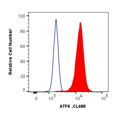
1X10^6 HeLa cells were intracellularly stained with 0.4 ug Anti-Human ATF6 and CoraLite®488-Conjugated AffiniPure Goat Anti-Mouse IgG(H+L) at dilution 1:1000 (red), or 0.4 ug Control Antibody. Cells were fixed and permeabilized with Transcription Factor Staining Buffer Kit.| Product Name | ATF6 Monoclonal antibody |
|---|---|
| Antibody Type | Primary Antibodies |
| Immunogen | Recombinant human ATF6 protein |
| Clonality | Monoclonal |
|---|---|
| Isotype | IgG1 |
| Host Species | Mouse |
| Tested Applications | FCICC/IFIHCWB |
| WB:1:5000-1:50000 IHC: ICC/IF1:250-1:1000:1:400-1:1600FC:0.40 ug per 10^6 cells in a 100 µl suspension |
|
| Species Reactivity | HumanMouseRat |
| Concentration | 1mg/ml |
| Purification | Protein G |
| Gene Symbol | ATF6 |
|---|---|
| Gene Synonyms | ACHM7 ATF6A ATP6alpha |
| Gene Full Name | activating transcription factor 6 |
| Gene Summary | This gene encodes a transcription factor that activates target genes for the unfolded protein response (UPR) during endoplasmic reticulum (ER) stress. Although it is a transcription factor, this protein is unusual in that it is synthesized as a transmembrane protein that is embedded in the ER. It functions as an ER stress sensor/transducer, and following ER stress-induced proteolysis, it functions as a nuclear transcription factor via a cis-acting ER stress response element (ERSE) that is present in the promoters of genes encoding ER chaperones. This protein has been identified as a survival factor for quiescent but not proliferative squamous carcinoma cells. There have been conflicting reports about the association of polymorphisms in this gene with diabetes in different populations, but another polymorphism has been associated with increased plasma cholesterol levels. This gene is also thought to be a potential therapeutic target for cystic fibrosis. [provided by RefSeq, Aug 2011] |
| Alternative Names | 3B7E4 activating transcription factor 6 ATF 6 ATF6 alpha ATF6A |
| Molecular Weight(MW) | 75 kDa(Observed band size: 90kDa-100 kDa) |
| Cellular Localization | Endoplasmic reticulum,Golgi apparatus,Membrane,Nucleus |
WB
Various lysates were subjected to SDS PAGE followed by western blot with ATF6 antibody at dilution of 1:10000 incubated at room temperature for 1.5 hours.
WB
WB result of ATF6 antibody (1:30000; incubated at room temperature for 1.5 hours) with sh-Control and sh-ATF6 transfected HEK-293 cells.
WB
WB result of ATF6 antibody (1:30000; incubated at room temperature for 1.5 hours) with sh-Control and sh-ATF6 transfected HeLa cells.
IHC
Immunohistochemical analysis of paraffin-embedded human breast cancer tissue slide using ATF6 antibody at dilution of 1:500 (under 10x lens. Heat mediated antigen retrieval with Tris-EDTA buffer (pH 9.0).
IHC
Immunohistochemical analysis of paraffin-embedded human cervical cancer tissue slide using ATF6 antibody at dilution of 1:500 (under 10x lens. Heat mediated antigen retrieval with Tris-EDTA buffer (pH 9.0).
IHC
Immunohistochemical analysis of paraffin-embedded human cervical cancer tissue slide using ATF6 antibody at dilution of 1:500 (under 40x lens. Heat mediated antigen retrieval with Tris-EDTA buffer (pH 9.0).
ICC/IF
Immunofluorescent analysis of (4% PFA) fixed HeLa cells using ATF6 antibody at dilution of 1:800 and Multi-rAb CoraLite ® Plus 488-Goat Anti-Mouse Recombinant Secondary Antibody (H+L), CL594-phalloidin (red).
FC
1X10^6 HeLa cells were intracellularly stained with 0.4 ug Anti-Human ATF6 and CoraLite®488-Conjugated AffiniPure Goat Anti-Mouse IgG(H+L) at dilution 1:1000 (red), or 0.4 ug Control Antibody. Cells were fixed and permeabilized with Transcription Factor Staining Buffer Kit.| Application Notes | WB:1:5000-1:50000 IHC: ICC/IF1:250-1:1000:1:400-1:1600FC:0.40 ug per 10^6 cells in a 100 µl suspension |
|---|
| Form | Liquid |
|---|---|
| Storage Instructions | Store at -20°C. Stable for one year after shipment. |
| Storage Buffer | PBS with 0.02% sodium azide and 50% glycerol pH 7.3. |


