WB
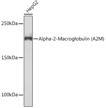
Western blot analysis of lysates from Hep G2 cells, using Alpha-2-Macroglobulin (Alpha-2-Macroglobulin(A2M)) Rabbit mAb at 1:1000 dilution.Secondary antibody: HRP-conjugated Goat anti-Rabbit IgG (H+L) at 1:10000 dilution.Lysates/proteins: 25μg per lane.Blocking buffer: 3% nonfat dry milk in TBST.Detection: ECL Basic Kit.Exposure time: 90s.IF-P
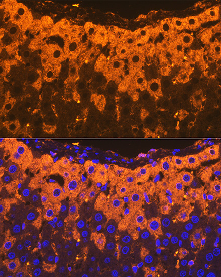
Immunofluorescence analysis of paraffinembedded rat liver using Alpha-2-Macroglobulin (Alpha-2-Macroglobulin (A2M))Rabbit mAb at dilution of 1:100 (40xlens). Secondary antibody: Cy3-conjugatedGoat anti-Rabbit IgG (H+L) at 1:500dilution. Blue: DAPI for nuclear staining.IF-P
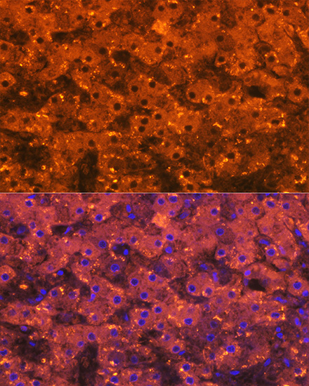
Immunofluorescence analysis of paraffinembedded human liver using Alpha-2-Macroglobulin (Alpha-2-Macroglobulin (A2M))Rabbit mAb at dilution of 1:100 (40xlens). Secondary antibody: Cy3-conjugatedGoat anti-Rabbit IgG (H+L) at 1:500dilution. Blue: DAPI for nuclear staining.IF-P
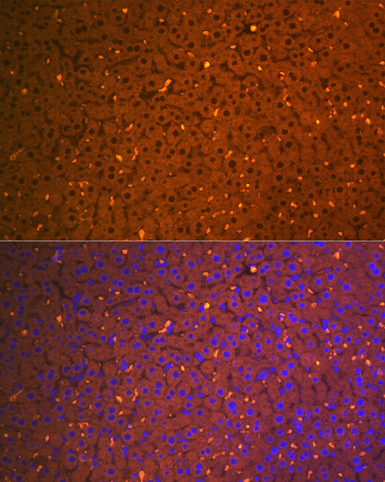
Immunofluorescence analysis of paraffinembedded mouse liver using Alpha-2-Macroglobulin (Alpha-2-Macroglobulin (A2M))Rabbit mAb at dilution of 1:100 (40xlens). Secondary antibody: Cy3-conjugatedGoat anti-Rabbit IgG (H+L) at 1:500dilution. Blue: DAPI for nuclear staining.IHC
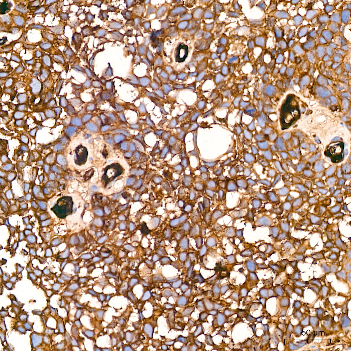
Immunohistochemistry analysis of paraffinembedded Human cervix cancer tissue usingAlpha-2-Macroglobulin (A2M) Rabbit mAb(A9752) at a dilution of 1:200 (40x lens).High pressure antigen retrieval performedwith 0.01M Citrate buffer (pH 6.0) prior toIHC staining.IHC
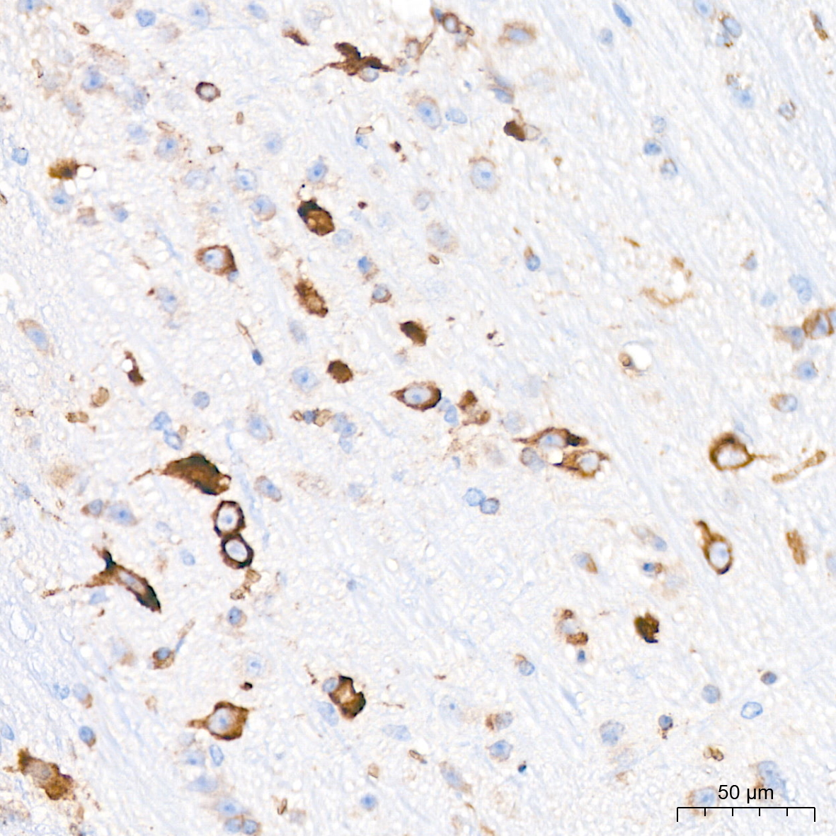
Immunohistochemistry analysis of paraffinembedded Mouse brain tissue using Alpha-2-Macroglobulin (A2M) Rabbit mAb ata dilution of 1:200 (40x lens). High pressureantigen retrieval performed with 0.01MCitrate buffer (pH 6.0) prior to IHC staining.IHC
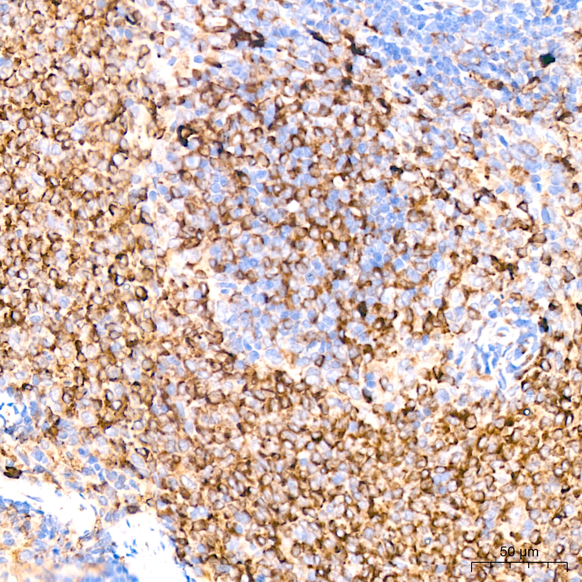
Immunohistochemistry analysis of paraffinembedded Rat spleen tissue using Alpha-2-Macroglobulin (A2M) Rabbit mAb ata dilution of 1:200 (40x lens). High pressureantigen retrieval performed with 0.01MCitrate buffer (pH 6.0) prior to IHC staining.| Product Name | Alpha-2-Macroglobulin (A2M) Rabbit mAb |
|---|---|
| Antibody Type | Primary Antibodies |
| Immunogen | Synthetic peptide. This information is considered to be commercially sensitive. |
| Clonality | Monoclonal |
|---|---|
| Isotype | IgG |
| Host Species | Rabbit |
| Tested Applications | IF-PIHCWB |
| WB:1:500-1:1000 IHC:1:200-1:2000 IF-P:1:100-1:400 |
|
| Species Reactivity | HumanMouseRat |
| Concentration | 1mg/ml |
| Purification | Affinity purified |
| Gene Symbol | A2M |
|---|---|
| Gene Synonyms | A2MD CPAMD5 FWP007 S863-7 |
| Gene Full Name | alpha-2-macroglobulin |
| Gene Summary | The protein encoded by this gene is a protease inhibitor and cytokine transporter. It uses a bait-and-trap mechanism to inhibit a broad spectrum of proteases, including trypsin, thrombin and collagenase. It can also inhibit inflammatory cytokines, and it thus disrupts inflammatory cascades. Mutations in this gene are a cause of alpha-2-macroglobulin deficiency. This gene is implicated in Alzheimer's disease (AD) due to its ability to mediate the clearance and degradation of A-beta, the major component of beta-amyloid deposits. A related pseudogene, which is also located on the p arm of chromosome 12, has been identified. [provided by RefSeq, Nov 2016] |
| Alternative Names | A2MD CPAMD5 FWP007 S863-7 Alpha-2-Macroglobulin (A2M) |
| Molecular Weight(MW) | 163kDa(Observed band size:185kDa) |
| Cellular Localization | Secreted. |
WB
Western blot analysis of lysates from Hep G2 cells, using Alpha-2-Macroglobulin (Alpha-2-Macroglobulin(A2M)) Rabbit mAb at 1:1000 dilution.Secondary antibody: HRP-conjugated Goat anti-Rabbit IgG (H+L) at 1:10000 dilution.Lysates/proteins: 25μg per lane.Blocking buffer: 3% nonfat dry milk in TBST.Detection: ECL Basic Kit.Exposure time: 90s.
IF-P
Immunofluorescence analysis of paraffinembedded rat liver using Alpha-2-Macroglobulin (Alpha-2-Macroglobulin (A2M))Rabbit mAb at dilution of 1:100 (40xlens). Secondary antibody: Cy3-conjugatedGoat anti-Rabbit IgG (H+L) at 1:500dilution. Blue: DAPI for nuclear staining.
IF-P
Immunofluorescence analysis of paraffinembedded human liver using Alpha-2-Macroglobulin (Alpha-2-Macroglobulin (A2M))Rabbit mAb at dilution of 1:100 (40xlens). Secondary antibody: Cy3-conjugatedGoat anti-Rabbit IgG (H+L) at 1:500dilution. Blue: DAPI for nuclear staining.
IF-P
Immunofluorescence analysis of paraffinembedded mouse liver using Alpha-2-Macroglobulin (Alpha-2-Macroglobulin (A2M))Rabbit mAb at dilution of 1:100 (40xlens). Secondary antibody: Cy3-conjugatedGoat anti-Rabbit IgG (H+L) at 1:500dilution. Blue: DAPI for nuclear staining.
IHC
Immunohistochemistry analysis of paraffinembedded Human cervix cancer tissue usingAlpha-2-Macroglobulin (A2M) Rabbit mAb(A9752) at a dilution of 1:200 (40x lens).High pressure antigen retrieval performedwith 0.01M Citrate buffer (pH 6.0) prior toIHC staining.
IHC
Immunohistochemistry analysis of paraffinembedded Mouse brain tissue using Alpha-2-Macroglobulin (A2M) Rabbit mAb ata dilution of 1:200 (40x lens). High pressureantigen retrieval performed with 0.01MCitrate buffer (pH 6.0) prior to IHC staining.
IHC
Immunohistochemistry analysis of paraffinembedded Rat spleen tissue using Alpha-2-Macroglobulin (A2M) Rabbit mAb ata dilution of 1:200 (40x lens). High pressureantigen retrieval performed with 0.01MCitrate buffer (pH 6.0) prior to IHC staining.| Application Notes | WB:1:500-1:1000 IHC:1:200-1:2000 IF-P:1:100-1:400 |
|---|
| Form | Liquid |
|---|---|
| Storage Instructions | Store at -20℃. Avoid freeze / thaw cycles. |
| Storage Buffer | PBS containing 50% glycerol and 0.05% BSA, preserved with proclin300 or sodium azide (as specified on the Certificate of Analysis), pH 7.3. |


