WB
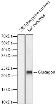
Western blot analysis of various lysates using Glucagon Rabbit mAb at 1:5000 dilutionincubated overnight at 4℃.Secondary antibody: HRP-conjugated Goat anti-Rabbit IgG (H+L) at 1:10000 dilution.Lysates/proteins: 25μg per lane.Blocking buffer: 3% nonfat dry milk in TBST.Detection: ECL Enhanced Kit.Exposure time: 180s.IHC
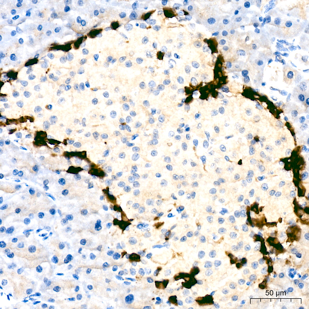
Immunohistochemistry analysis of paraffinembedded Human pancreas tissue usingGlucagon Rabbit mAb at a dilutionof 1:35000 (40x lens). High pressure antigenretrieval performed with 0.01M Tris-EDTABuffer (pH 9.0) prior to IHC staining.IF-P
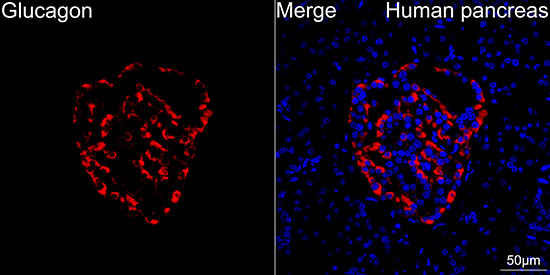
Confocal imaging of paraffin-embeddedHuman pancreas tissue using GlucagonRabbit mAb (dilution 1:1500)followed by a further incubation with Cy3Goat Anti-Rabbit IgG (H+L) (dilution1:500) (Red). DAPI was used for nuclearstaining (Blue). High pressure antigenretrieval performed with 0.01M Citrate Buffer(pH 6.0) prior to IF staining. Objective: 40x.IF-P
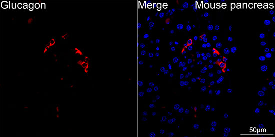
Confocal imaging of paraffin-embeddedMouse pancreas tissue using GlucagonRabbit mAb (dilution 1:1500)followed by a further incubation with Cy3Goat Anti-Rabbit IgG (H+L) (dilution1:500) (Red). DAPI was used for nuclearstaining (Blue). High pressure antigenretrieval performed with 0.01M Citrate Buffer(pH 6.0) prior to IF staining. Objective: 40x.IF-P
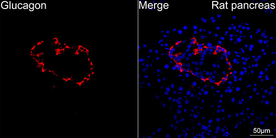
Confocal imaging of paraffin-embedded Ratpancreas tissue using Glucagon Rabbit mAb(dilution 1:1500) followed by afurther incubation with Cy3 Goat Anti-RabbitIgG (H+L) (dilution 1:500) (Red).DAPI was used for nuclear staining (Blue).High pressure antigen retrieval performedwith 0.01M Citrate Buffer (pH 6.0) prior to IFstaining. Objective: 40x.| Product Name | Glucagon Rabbit mAb |
|---|---|
| Antibody Type | Primary Antibodies |
| Immunogen | Synthetic peptide. This information is considered to be commercially sensitive. |
| Clonality | Monoclonal |
|---|---|
| Isotype | IgG |
| Host Species | Rabbit |
| Tested Applications | IF-PIHCWB |
| WB:1:2000-1:6000 IHC:1:10000-1:40000 IF-P:1:1000-1:5000 |
|
| Species Reactivity | HumanMouseRat |
| Concentration | 1mg/ml |
| Purification | Affinity purified |
| Gene Symbol | GCG |
|---|---|
| Gene Synonyms | GLP1 GLP2 GRPP GLP-1 |
| Gene Full Name | glucagon |
| Gene Summary | The protein encoded by this gene is actually a preproprotein that is cleaved into four distinct mature peptides. One of these, glucagon, is a pancreatic hormone that counteracts the glucose-lowering action of insulin by stimulating glycogenolysis and gluconeogenesis. Glucagon is a ligand for a specific G-protein linked receptor whose signalling pathway controls cell proliferation. Two of the other peptides are secreted from gut endocrine cells and promote nutrient absorption through distinct mechanisms. Finally, the fourth peptide is similar to glicentin, an active enteroglucagon. [provided by RefSeq, Jul 2008] |
| Alternative Names | GLP1 GLP2 GRPP GLP-1 Glucagon |
| Molecular Weight(MW) | 21kDa(Observed band size:21kDa) |
| Cellular Localization | Secreted. |
WB
Western blot analysis of various lysates using Glucagon Rabbit mAb at 1:5000 dilutionincubated overnight at 4℃.Secondary antibody: HRP-conjugated Goat anti-Rabbit IgG (H+L) at 1:10000 dilution.Lysates/proteins: 25μg per lane.Blocking buffer: 3% nonfat dry milk in TBST.Detection: ECL Enhanced Kit.Exposure time: 180s.
IHC
Immunohistochemistry analysis of paraffinembedded Human pancreas tissue usingGlucagon Rabbit mAb at a dilutionof 1:35000 (40x lens). High pressure antigenretrieval performed with 0.01M Tris-EDTABuffer (pH 9.0) prior to IHC staining.
IF-P
Confocal imaging of paraffin-embeddedHuman pancreas tissue using GlucagonRabbit mAb (dilution 1:1500)followed by a further incubation with Cy3Goat Anti-Rabbit IgG (H+L) (dilution1:500) (Red). DAPI was used for nuclearstaining (Blue). High pressure antigenretrieval performed with 0.01M Citrate Buffer(pH 6.0) prior to IF staining. Objective: 40x.
IF-P
Confocal imaging of paraffin-embeddedMouse pancreas tissue using GlucagonRabbit mAb (dilution 1:1500)followed by a further incubation with Cy3Goat Anti-Rabbit IgG (H+L) (dilution1:500) (Red). DAPI was used for nuclearstaining (Blue). High pressure antigenretrieval performed with 0.01M Citrate Buffer(pH 6.0) prior to IF staining. Objective: 40x.
IF-P
Confocal imaging of paraffin-embedded Ratpancreas tissue using Glucagon Rabbit mAb(dilution 1:1500) followed by afurther incubation with Cy3 Goat Anti-RabbitIgG (H+L) (dilution 1:500) (Red).DAPI was used for nuclear staining (Blue).High pressure antigen retrieval performedwith 0.01M Citrate Buffer (pH 6.0) prior to IFstaining. Objective: 40x.| Application Notes | WB:1:2000-1:6000 IHC:1:10000-1:40000 IF-P:1:1000-1:5000 |
|---|
| Form | Liquid |
|---|---|
| Storage Instructions | Store at -20℃. Avoid freeze / thaw cycles. |
| Storage Buffer | PBS containing 50% glycerol and 0.05% BSA, preserved with proclin300 or sodium azide (as specified on the Certificate of Analysis), pH 7.3. |


