WB
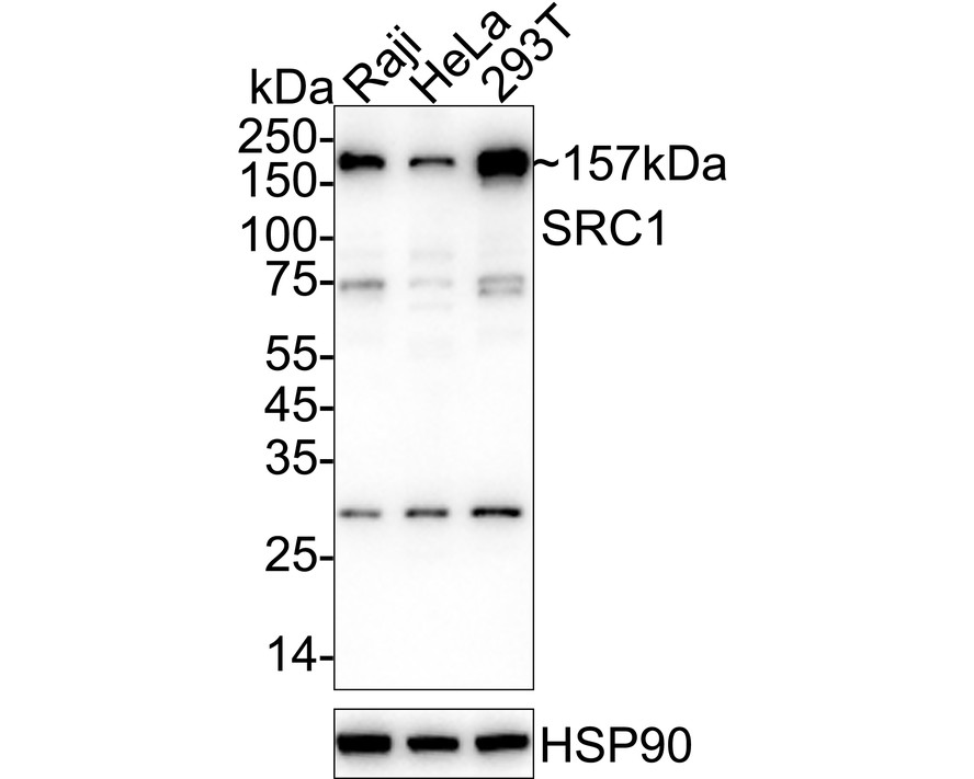
Western blot analysis of SRC1 on different lysates with Rabbit anti-SRC1 antibody at 1/2000 dilution. Lane 1: Raji cell lysate, Lane 2: HeLa cell lysate, Lane 3: 293T cell lysate, Lysates/proteins at 20 µg/Lane. Exposure time: 1 minute; 4-20% SDS-PAGE gel. Proteins were transferred to a PVDF membrane and blocked with 5% NFDM/TBST for 1 hour at room temperature. The primary antibody at 1/2000 dilution was used in 5% NFDM/TBST at 4℃ overnight. Goat Anti-Rabbit IgG - HRP Secondary Antibody at 1/50000 dilution was used for 1 hour at room temperature.ICC/IF
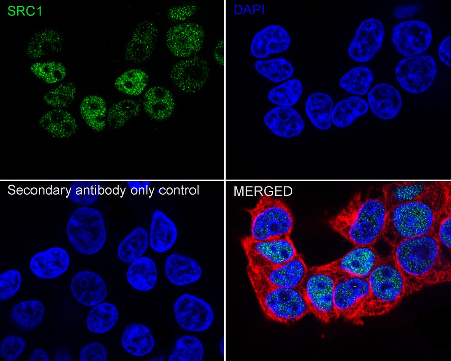
Immunocytochemistry analysis of 293T cells labeling SRC1 with Rabbit anti-SRC1 antibody at 1/100 dilution. Cells were fixed in 4% paraformaldehyde for 15 minutes at room temperature, permeabilized with 0.1% Triton X-100 in PBS for 15 minutes at room temperature, then blocked with 1% BSA in 10% negative goat serum for 1 hour at room temperature. Cells were then incubated with Rabbit anti-SRC1 antibody at 1/100 dilution in 1% BSA in PBST overnight at 4 ℃. Goat Anti-Rabbit IgG H&L (488) was used as the secondary antibody at 1/1,000 dilution. PBS instead of the primary antibody was used as the secondary antibody only control. Nuclear DNA was labelled in blue with DAPI. Beta tubulin (red) was stained at 1/100 dilution overnight at +4℃. Goat Anti-Mouse IgG H&L ( 594) was used as the secondary antibody at 1/1000 dilution.IHC
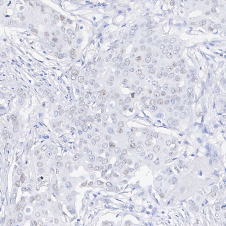
Immunohistochemical analysis of paraffin-embedded human breast cancer tissue with Rabbit anti-SRC1 antibody at 1/1000 dilution. The section was pre-treated using heat mediated antigen retrieval with sodium citrate buffer (pH 6.0) (high pressure) for 2 minutes. The tissues were blocked in 1% BSA for 20 minutes at room temperature, washed with ddH2O and PBS, and then probed with the primary antibody at 1/1000 dilution for 1 hour at room temperature. The detection was performed using an HRP conjugated compact polymer system. DAB was used as the chromogen. Tissues were counterstained with hematoxylin and mounted with DPX.| Product Name | SRC1 Recombinant Rabbit Monoclonal Antibody |
|---|---|
| Antibody Type | Primary Antibodies |
| Immunogen | Recombinant protein within Human SRC1 aa 380-770 / 1,441. |
| Clonality | Monoclonal |
|---|---|
| Isotype | IgG |
| Host Species | Rabbit |
| Tested Applications | ICC/IFIHCWB |
| WB:1:500-1:2000 ICC/IF:1:100 IHC:1:500-1:1000 |
|
| Species Reactivity | Human |
| Concentration | 1mg/ml |
| Purification | Affinity purified |
| Gene Symbol | NCOA1 |
|---|---|
| Gene Synonyms | SRC1 KAT13A RIP160 F-SRC-1 bHLHe42 bHLHe74 |
| Gene Full Name | nuclear receptor coactivator 1 |
| Gene Summary | The protein encoded by this gene acts as a transcriptional coactivator for steroid and nuclear hormone receptors. It is a member of the p160/steroid receptor coactivator (SRC) family and like other family members has histone acetyltransferase activity and contains a nuclear localization signal, as well as bHLH and PAS domains. The product of this gene binds nuclear receptors directly and stimulates the transcriptional activities in a hormone-dependent fashion. Alternatively spliced transcript variants encoding different isoforms have been identified. [provided by RefSeq, Jul 2008] |
| Alternative Names | bHLHe74 antibody Class E basic helix-loop-helix protein 74 antibody F SRC 1 antibody Hin 2 protein antibody Hin2 protein antibody MGC129719 antibody MGC129720 antibody mNRC 1 antibody NCoA 1 antibody NCoA-1 antibody bHLHe74 antibody Class E basic helix-loop-helix protein 74 antibody F SRC 1 antibody Hin 2 protein antibody Hin2 protein antibody MGC129719 antibody MGC129720 antibody mNRC 1 antibody NCoA 1 antibody NCoA-1 antibody Ncoa1 antibody NCOA1_HUMAN antibody Nuclear receptor coactivator 1 antibody Nuclear receptor coactivator protein 1 antibody NY REN 52 antigen antibody Protein Hin 2 antibody Protein Hin-2 antibody Protein Hin2 antibody Renal carcinoma antigen NY REN 52 antibody Renal carcinoma antigen NY-REN-52 antibody RIP 160 antibody RIP160 antibody SRC 1 antibody SRC-1 antibody Steroid receptor coactivator 1 antibody |
| Molecular Weight(MW) | 157kDa(Observed band size:157kDa) |
| Cellular Localization | Nucleus. |
WB
Western blot analysis of SRC1 on different lysates with Rabbit anti-SRC1 antibody at 1/2000 dilution. Lane 1: Raji cell lysate, Lane 2: HeLa cell lysate, Lane 3: 293T cell lysate, Lysates/proteins at 20 µg/Lane. Exposure time: 1 minute; 4-20% SDS-PAGE gel. Proteins were transferred to a PVDF membrane and blocked with 5% NFDM/TBST for 1 hour at room temperature. The primary antibody at 1/2000 dilution was used in 5% NFDM/TBST at 4℃ overnight. Goat Anti-Rabbit IgG - HRP Secondary Antibody at 1/50000 dilution was used for 1 hour at room temperature.
ICC/IF
Immunocytochemistry analysis of 293T cells labeling SRC1 with Rabbit anti-SRC1 antibody at 1/100 dilution. Cells were fixed in 4% paraformaldehyde for 15 minutes at room temperature, permeabilized with 0.1% Triton X-100 in PBS for 15 minutes at room temperature, then blocked with 1% BSA in 10% negative goat serum for 1 hour at room temperature. Cells were then incubated with Rabbit anti-SRC1 antibody at 1/100 dilution in 1% BSA in PBST overnight at 4 ℃. Goat Anti-Rabbit IgG H&L (488) was used as the secondary antibody at 1/1,000 dilution. PBS instead of the primary antibody was used as the secondary antibody only control. Nuclear DNA was labelled in blue with DAPI. Beta tubulin (red) was stained at 1/100 dilution overnight at +4℃. Goat Anti-Mouse IgG H&L ( 594) was used as the secondary antibody at 1/1000 dilution.
IHC
Immunohistochemical analysis of paraffin-embedded human breast cancer tissue with Rabbit anti-SRC1 antibody at 1/1000 dilution. The section was pre-treated using heat mediated antigen retrieval with sodium citrate buffer (pH 6.0) (high pressure) for 2 minutes. The tissues were blocked in 1% BSA for 20 minutes at room temperature, washed with ddH2O and PBS, and then probed with the primary antibody at 1/1000 dilution for 1 hour at room temperature. The detection was performed using an HRP conjugated compact polymer system. DAB was used as the chromogen. Tissues were counterstained with hematoxylin and mounted with DPX.| Application Notes | WB:1:500-1:2000 ICC/IF:1:100 IHC:1:500-1:1000 |
|---|
| Form | Liquid |
|---|---|
| Storage Instructions | Shipped at 4℃. Store at +4℃ short term (1-2 weeks). It is recommended to aliquot into single-use upon delivery. Store at -20℃ long term. |
| Storage Buffer | 1*TBS (pH7.4), 0.05% BSA, 40% Glycerol. Preservative: 0.05% Sodium Azide. |


