WB
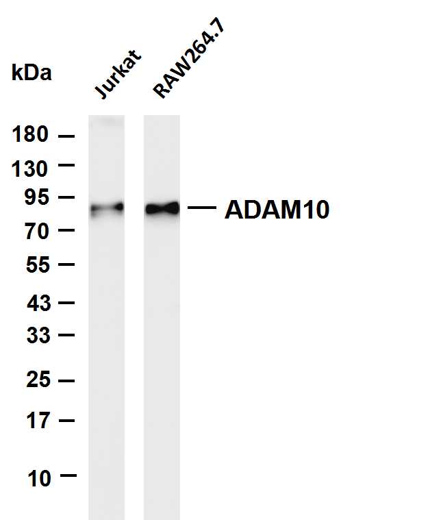
Various whole cell lysates were separated by 4-20% SDS-PAGE, and the membrane was blotted with anti-ADAM10 antibody. The HRP-conjugated Goat anti-Rabbit IgG(H + L) antibody was used to detect the antibody. Lane 1: Jurkat, Lane 2: RAW264.7.ICC/IF
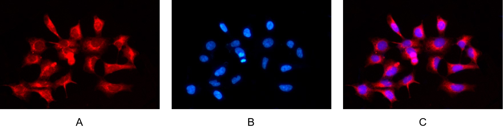
Immunofluorescence analysis of HEK293. Picture A: ADAM10 antibody (red). Picture B: DAPI (blue). Picture C: Merge of A+B.IHC
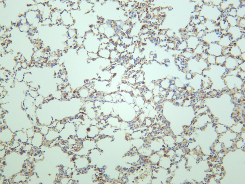
Mouse lung was stained with anti-ADAM10 rabbit antibody.IHC
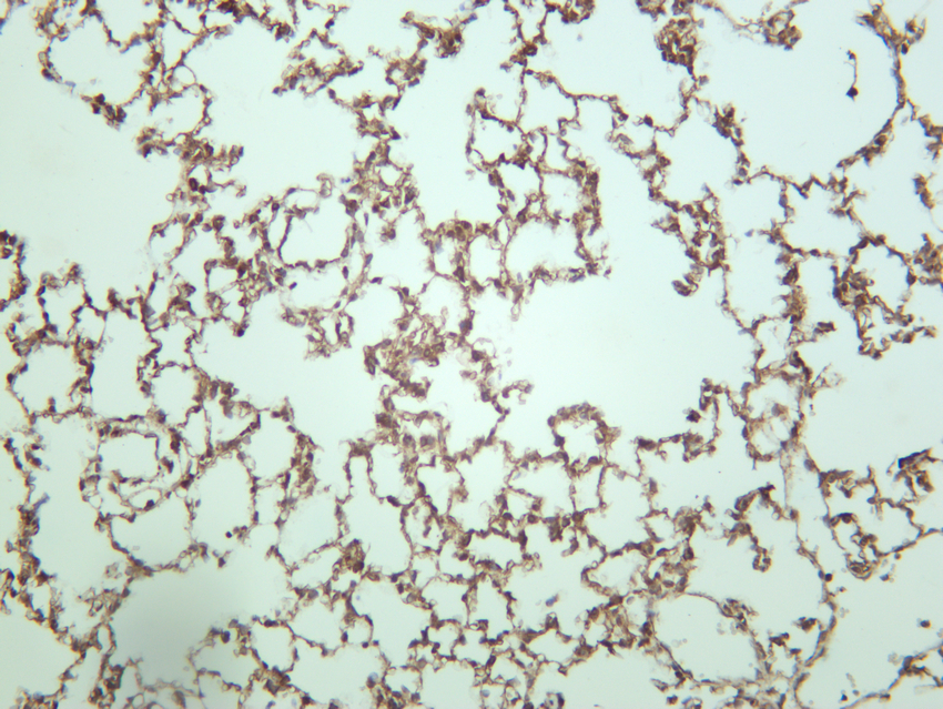
Rat lung was stained with anti-ADAM10 rabbit antibody.IHC
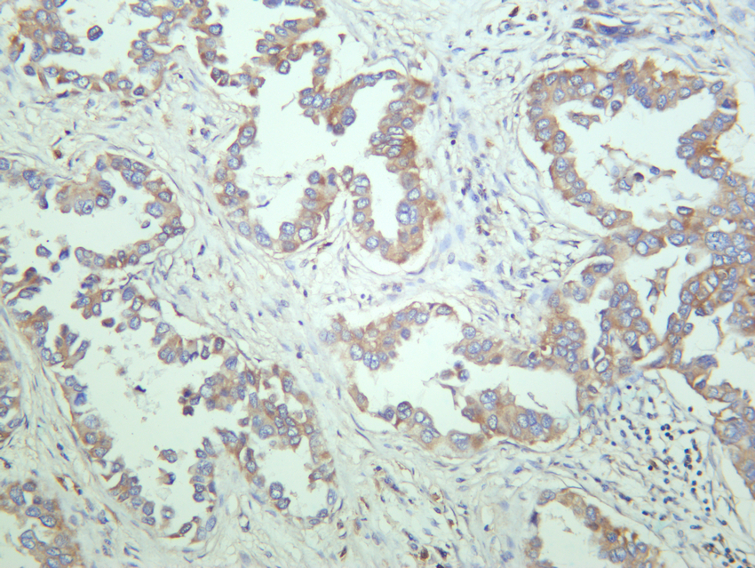
Human lung carcinoma was stained with anti-ADAM10 rabbit antibody.| Product Name | ADAM10 Rabbit mAb |
|---|---|
| Antibody Type | Primary Antibodies |
| Clonality | monoclonal |
|---|---|
| Isotype | IgG,K |
| Host Species | Rabbit |
| Tested Applications | ICC/IFIHCWB |
| WB:1:2000-1:10000 IHC:1:200-1:2000 ICC/IF:1:200-1:1000 |
|
| Species Reactivity | HumanMouseRat |
| Concentration | 1mg/ml |
| Purification | Protein A |
| Gene Symbol | ADAM10 |
|---|---|
| Gene Synonyms | RAK kuz AD10 AD18 MADM CD156c CDw156 HsT18717 |
| Gene Full Name | ADAM metallopeptidase domain 10 |
| Gene Summary | Members of the ADAM family are cell surface proteins with a unique structure possessing both potential adhesion and protease domains. This gene encodes and ADAM family member that cleaves many proteins including TNF-alpha and E-cadherin. Alternate splicing results in multiple transcript variants encoding different proteins that may undergo similar processing. [provided by RefSeq, Feb 2016] |
| Alternative Names | ADAM10 KUZ MADM Disintegrin and metalloproteinase domain-containing protein 10 ADAM 10 CDw156 Kuzbanian protein homolog Mammalian disintegrin-metalloprotease CD156c |
| Molecular Weight(MW) | 84kDa(Observed MW 84kDa) |
| Cellular Localization | Cell membrane ; Single-pass type I membrane protein . Golgi apparatus membrane ; Single-pass type I membrane protein . Cytoplasmic vesicle, clathrin-coated vesicle . Cell projection, axon . Cell projection, dendrite . Cell junction, adherens junction . Cytoplasm . |
WB
Various whole cell lysates were separated by 4-20% SDS-PAGE, and the membrane was blotted with anti-ADAM10 antibody. The HRP-conjugated Goat anti-Rabbit IgG(H + L) antibody was used to detect the antibody. Lane 1: Jurkat, Lane 2: RAW264.7.
ICC/IF
Immunofluorescence analysis of HEK293. Picture A: ADAM10 antibody (red). Picture B: DAPI (blue). Picture C: Merge of A+B.
IHC
Mouse lung was stained with anti-ADAM10 rabbit antibody.
IHC
Rat lung was stained with anti-ADAM10 rabbit antibody.
IHC
Human lung carcinoma was stained with anti-ADAM10 rabbit antibody.| Application Notes | WB:1:2000-1:10000 IHC:1:200-1:2000 ICC/IF:1:200-1:1000 |
|---|
| Form | Liquid |
|---|---|
| Storage Instructions | -15°C to -25°C/1 year(Do not lower than -25°C) |
| Storage Buffer | PBS, 50% glycerol, 0.05% Proclin 300, 0.05%BSA. |


