WB
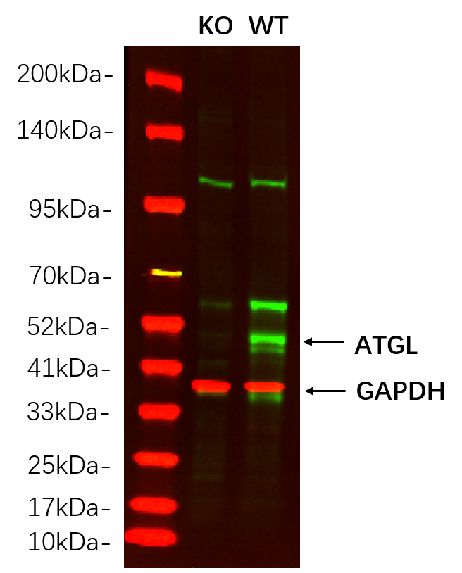
Western blot analysis of lysates from HAP1 WT and knockout cell. (Green) primary antibody was diluted at 1:5000, 4°over night, Dylight 800 secondary antibody was diluted at 1:10000, 37° 1hour. (Red) GAPDH Monoclonal Antibody(5B7) antibody was diluted at 1:5000 as loading control, 4° over night, Dylight 680 secondary antibody was diluted at 1:10000, 37° 1hour.WB
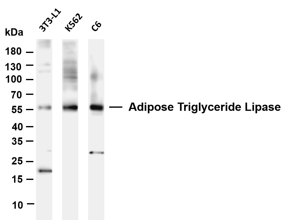
Various whole cell lysates were separated by 4-20% SDS-PAGE, and the membrane was blotted with anti- ATGL antibody. The HRP-conjugated Goat anti-Rabbit IgG(H + L) antibody was used to detect the antibody. Lane 1: 3T3-L1 Lane 2: K562 Lane 3: C6 .IHC
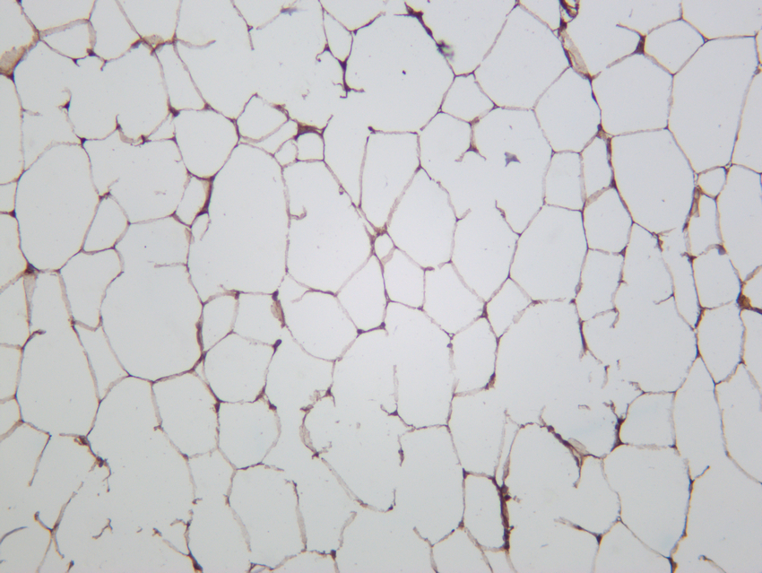
Rat white adipose was stained with anti-ATGL rabbit antibody.| Product Name | ATGL Rabbit mAb |
|---|---|
| Antibody Type | Primary Antibodies |
| Clonality | monoclonal |
|---|---|
| Isotype | IgG,K |
| Host Species | Rabbit |
| Tested Applications | IHCWB |
| WB:1:2000-1:10000 IHC:1:200-1:1000 |
|
| Species Reactivity | HumanMouseRat |
| Concentration | 1mg/ml |
| Purification | Protein A |
| Gene Symbol | PNPLA2 |
|---|---|
| Gene Synonyms | ATGL TTS2 PEDF-R FP17548 TTS-2.2 iPLA2zeta 1110001C14Rik |
| Gene Full Name | patatin like domain 2, triacylglycerol lipase |
| Gene Summary | This gene encodes an enzyme which catalyzes the first step in the hydrolysis of triglycerides in adipose tissue. Mutations in this gene are associated with neutral lipid storage disease with myopathy. [provided by RefSeq, Jul 2010] |
| Molecular Weight(MW) | 55kDa(Observed MW 55kDa) |
| Cellular Localization | Lipid droplet . Cell membrane ; Multi-pass membrane protein . Cytoplasm . |
WB
Western blot analysis of lysates from HAP1 WT and knockout cell. (Green) primary antibody was diluted at 1:5000, 4°over night, Dylight 800 secondary antibody was diluted at 1:10000, 37° 1hour. (Red) GAPDH Monoclonal Antibody(5B7) antibody was diluted at 1:5000 as loading control, 4° over night, Dylight 680 secondary antibody was diluted at 1:10000, 37° 1hour.
WB
Various whole cell lysates were separated by 4-20% SDS-PAGE, and the membrane was blotted with anti- ATGL antibody. The HRP-conjugated Goat anti-Rabbit IgG(H + L) antibody was used to detect the antibody. Lane 1: 3T3-L1 Lane 2: K562 Lane 3: C6 .
IHC
Rat white adipose was stained with anti-ATGL rabbit antibody.| Application Notes | WB:1:2000-1:10000 IHC:1:200-1:1000 |
|---|
| Form | Liquid |
|---|---|
| Storage Instructions | -15°C to -25°C/1 year(Do not lower than -25°C) |
| Storage Buffer | PBS, 50% glycerol, 0.05% Proclin 300, 0.05%BSA |


