WB
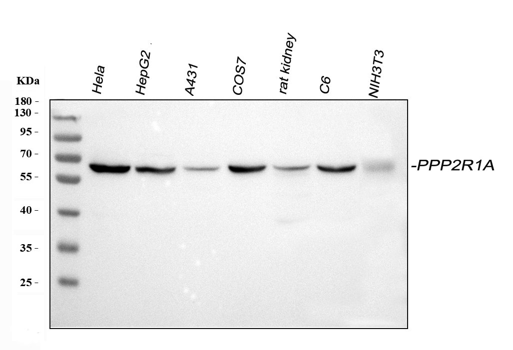
Western blot analysis of PPP2R1A using anti-PPP2R1A antibody. The sample well of each lane was loaded with 30 ug of sample under reducing conditions. Lane 1: human Hela whole cell lysates, Lane 2: human HepG2 whole cell lysates, Lane 3: human A431 whole cell lysates, Lane 4: monkey COS7 whole cell lysates, Lane 5: rat kidney tissue lysates, Lane 6: rat C6 whole cell lysates, Lane 7: mouse NIH/3T3 whole cell lysates. After electrophoresis, proteins were transferred to a membrane. Then the membrane was incubated with rabbit anti-PPP2R1A antigen affinity purified polyclonal antibody at a dilution of 1:1000 and probed with a goat anti-rabbit IgG-HRP secondary antibody. The signal is developed using ECL Plus Western Blotting Substrate.IHC
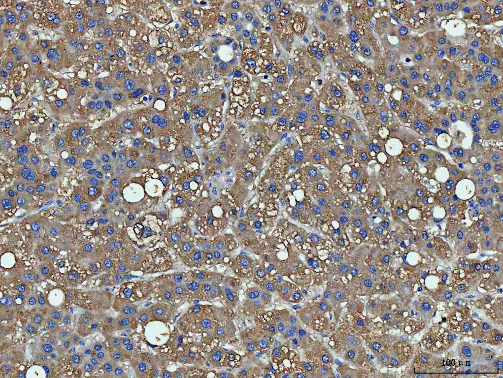
IHC analysis of PPP2R1A using anti-PPP2R1A antibody. PPP2R1A was detected in a paraffin-embedded section of human liver cancer tissue. The tissue section was incubated with rabbit anti-PPP2R1A Antibody at a dilution of 1:200 and developed using HRP Conjugated Rabbit IgG Super Vision Assay Kit with DAB as the chromogen.ICC/IF
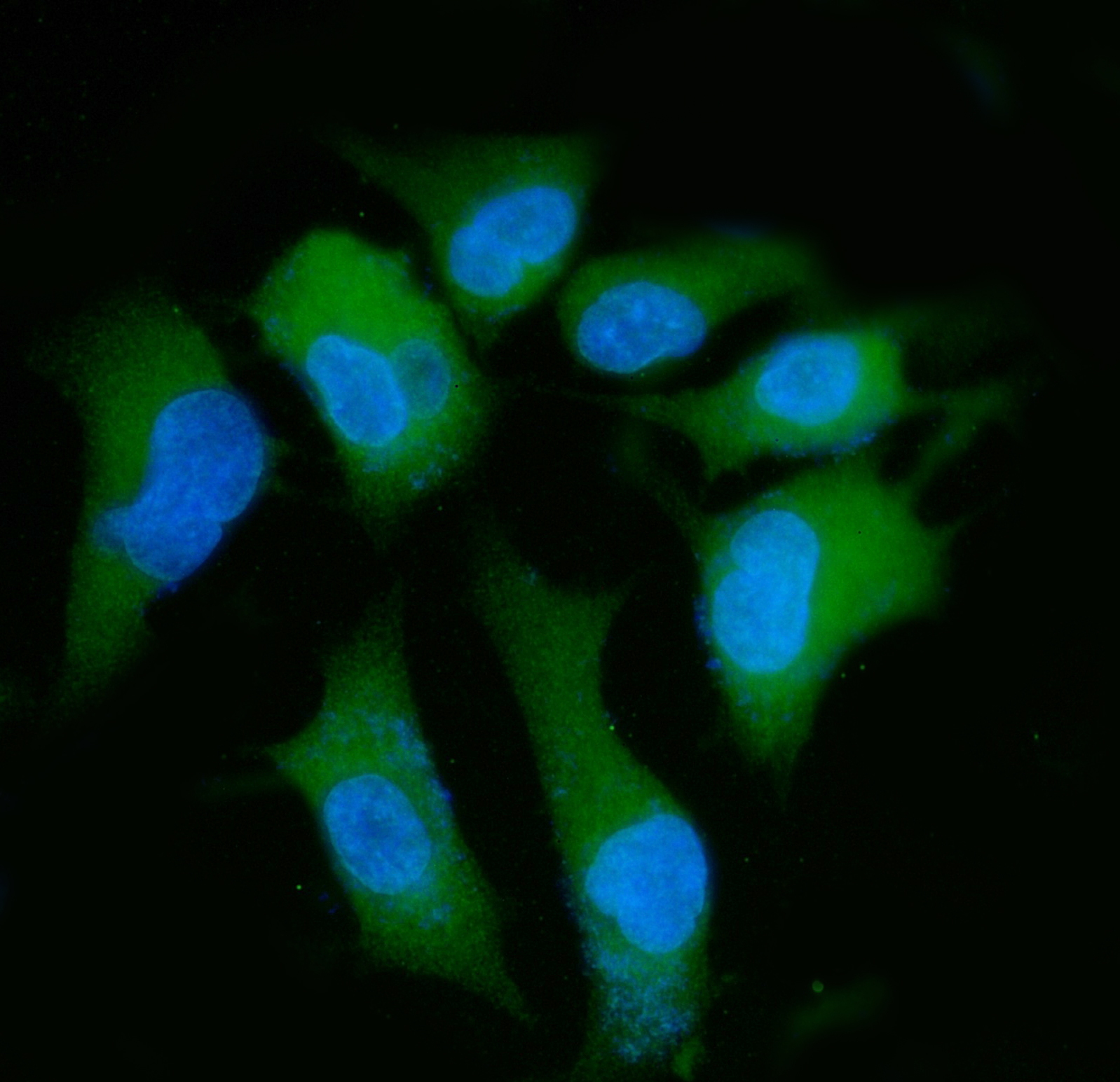
ICC/IF analysis of PPP2R1A using anti-PPP2R1A antibody. PPP2R1A was detected in an immunocytochemical section of A549 cells. The section was incubated with rabbit anti-PPP2R1A Antibody at a dilution of 1:100. Fluoro488 Conjugated Goat Anti-Rabbit IgG (Green) was used as secondary antibody. The section was counterstained with DAPI (Blue).FC
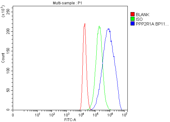
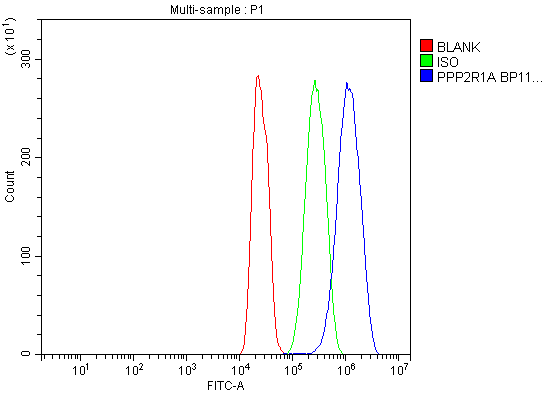
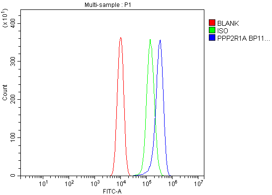
Flow Cytometry analysis of 293T cells using anti-PPP2R1A antibody. Overlay histogram showing 293T cells stained with anti-PPP2R1A antibody (Blue line). To facilitate intracellular staining, cells were fixed with 4% paraformaldehyde and permeabilized with permeabilization buffer. The cells were blocked with 10% normal goat serum. And then incubated with rabbit anti-PPP2R1A Antibody at 1:100 dilution for 30 min at 20°C. Fluoro488 conjugated goat anti-rabbit IgG was used as secondary antibody at 1:100 dilution for 30 minutes at 20°C. Isotype control antibody (Green line) was rabbit IgG at 1:100 dilution used under the same conditions. Unlabelled sample without incubation with primary antibody and secondary antibody (Red line) was used as a blank control.FC
Flow Cytometry analysis of C6 cells using anti-PPP2R1A antibody. Overlay histogram showing C6 cells stained with anti-PPP2R1A antibody (Blue line). To facilitate intracellular staining, cells were fixed with 4% paraformaldehyde and permeabilized with permeabilization buffer. The cells were blocked with 10% normal goat serum. And then incubated with rabbit anti-PPP2R1A Antibody at 1:100 dilution for 30 min at 20°C. Fluoro488 conjugated goat anti-rabbit IgG was used as secondary antibody at 1:100 dilution for 30 minutes at 20°C. Isotype control antibody (Green line) was rabbit IgG at 1:100 dilution used under the same conditions. Unlabelled sample without incubation with primary antibody and secondary antibody (Red line) was used as a blank control.FC
Flow Cytometry analysis of Raw264.7 cells using anti-PPP2R1A antibody. Overlay histogram showing Raw264.7 cells stained with anti-PPP2R1A antibody (Blue line). To facilitate intracellular staining, cells were fixed with 4% paraformaldehyde and permeabilized with permeabilization buffer. The cells were blocked with 10% normal goat serum. And then incubated with rabbit anti-PPP2R1A Antibody at 1:100 dilution for 30 min at 20°C. Fluoro488 conjugated goat anti-rabbit IgG was used as secondary antibody at 1:100 dilution for 30 minutes at 20°C. Isotype control antibody (Green line) was rabbit IgG at 1:100 dilution used under the same conditions. Unlabelled sample without incubation with primary antibody and secondary antibody (Red line) was used as a blank control.| Product Name | Anti-PPP2R1A Antibody |
|---|---|
| Antibody Type | Primary Antibodies |
| Immunogen | A synthetic peptide corresponding to a sequence in the middle region of human PPP2R1A, which shares 95.2% amino acid (aa) sequence identity with mouse PPP2R1A, respectively. |
| Clonality | Polyclonal |
|---|---|
| Isotype | IgG |
| Host Species | Rabbit |
| Tested Applications | FCICC/IFIHCWB |
| WB:1:500-2000 IHC:1:50-400 ICC/IF:1:50-400 FC:1:50-200 |
|
| Species Reactivity | HumanMonkeyMouseRat |
| Concentration | 1mg/ml |
| Purification | Affinity purified |
| Gene Symbol | PPP2R1A |
|---|---|
| Gene Synonyms | HJS2 MRD36 PP2AA PR65A PP2AAALPHA PP2A-Aalpha |
| Gene Full Name | protein phosphatase 2 scaffold subunit Aalpha |
| Gene Summary | This gene encodes a constant regulatory subunit of protein phosphatase 2. Protein phosphatase 2 is one of the four major Ser/Thr phosphatases, and it is implicated in the negative control of cell growth and division. It consists of a common heteromeric core enzyme, which is composed of a catalytic subunit and a constant regulatory subunit, that associates with a variety of regulatory subunits. The constant regulatory subunit A serves as a scaffolding molecule to coordinate the assembly of the catalytic subunit and a variable regulatory B subunit. This gene encodes an alpha isoform of the constant regulatory subunit A. Alternatively spliced transcript variants have been described. [provided by RefSeq, Apr 2010] |
| Alternative Names | PPP2R1A PR65A |
| Molecular Weight(MW) | 65kDa(Observed MW 65kDa) |
| Cellular Localization | Cell membrane |
WB
Western blot analysis of PPP2R1A using anti-PPP2R1A antibody. The sample well of each lane was loaded with 30 ug of sample under reducing conditions. Lane 1: human Hela whole cell lysates, Lane 2: human HepG2 whole cell lysates, Lane 3: human A431 whole cell lysates, Lane 4: monkey COS7 whole cell lysates, Lane 5: rat kidney tissue lysates, Lane 6: rat C6 whole cell lysates, Lane 7: mouse NIH/3T3 whole cell lysates. After electrophoresis, proteins were transferred to a membrane. Then the membrane was incubated with rabbit anti-PPP2R1A antigen affinity purified polyclonal antibody at a dilution of 1:1000 and probed with a goat anti-rabbit IgG-HRP secondary antibody. The signal is developed using ECL Plus Western Blotting Substrate.
IHC
IHC analysis of PPP2R1A using anti-PPP2R1A antibody. PPP2R1A was detected in a paraffin-embedded section of human liver cancer tissue. The tissue section was incubated with rabbit anti-PPP2R1A Antibody at a dilution of 1:200 and developed using HRP Conjugated Rabbit IgG Super Vision Assay Kit with DAB as the chromogen.
ICC/IF
ICC/IF analysis of PPP2R1A using anti-PPP2R1A antibody. PPP2R1A was detected in an immunocytochemical section of A549 cells. The section was incubated with rabbit anti-PPP2R1A Antibody at a dilution of 1:100. Fluoro488 Conjugated Goat Anti-Rabbit IgG (Green) was used as secondary antibody. The section was counterstained with DAPI (Blue).
FC
Flow Cytometry analysis of 293T cells using anti-PPP2R1A antibody. Overlay histogram showing 293T cells stained with anti-PPP2R1A antibody (Blue line). To facilitate intracellular staining, cells were fixed with 4% paraformaldehyde and permeabilized with permeabilization buffer. The cells were blocked with 10% normal goat serum. And then incubated with rabbit anti-PPP2R1A Antibody at 1:100 dilution for 30 min at 20°C. Fluoro488 conjugated goat anti-rabbit IgG was used as secondary antibody at 1:100 dilution for 30 minutes at 20°C. Isotype control antibody (Green line) was rabbit IgG at 1:100 dilution used under the same conditions. Unlabelled sample without incubation with primary antibody and secondary antibody (Red line) was used as a blank control.
FC
Flow Cytometry analysis of C6 cells using anti-PPP2R1A antibody. Overlay histogram showing C6 cells stained with anti-PPP2R1A antibody (Blue line). To facilitate intracellular staining, cells were fixed with 4% paraformaldehyde and permeabilized with permeabilization buffer. The cells were blocked with 10% normal goat serum. And then incubated with rabbit anti-PPP2R1A Antibody at 1:100 dilution for 30 min at 20°C. Fluoro488 conjugated goat anti-rabbit IgG was used as secondary antibody at 1:100 dilution for 30 minutes at 20°C. Isotype control antibody (Green line) was rabbit IgG at 1:100 dilution used under the same conditions. Unlabelled sample without incubation with primary antibody and secondary antibody (Red line) was used as a blank control.
FC
Flow Cytometry analysis of Raw264.7 cells using anti-PPP2R1A antibody. Overlay histogram showing Raw264.7 cells stained with anti-PPP2R1A antibody (Blue line). To facilitate intracellular staining, cells were fixed with 4% paraformaldehyde and permeabilized with permeabilization buffer. The cells were blocked with 10% normal goat serum. And then incubated with rabbit anti-PPP2R1A Antibody at 1:100 dilution for 30 min at 20°C. Fluoro488 conjugated goat anti-rabbit IgG was used as secondary antibody at 1:100 dilution for 30 minutes at 20°C. Isotype control antibody (Green line) was rabbit IgG at 1:100 dilution used under the same conditions. Unlabelled sample without incubation with primary antibody and secondary antibody (Red line) was used as a blank control.| Application Notes | WB:1:500-2000 IHC:1:50-400 ICC/IF:1:50-400 FC:1:50-200 |
|---|
| Form | Liquid |
|---|---|
| Storage Instructions | 12 months from date of receipt,-20℃ as supplied. |
| Storage Buffer | 500 ug/ml antibody with PBS, 0.02% NaN3, 1 mg/ml BSA and 50% glycerol. |


