WB
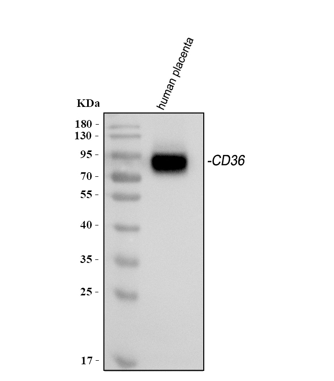
Western blot analysis of anti-CD36 antibody. The sample well of each lane was loaded with 30 ug of sample under reducing conditions. Lane 1: human placenta tissue lysates. After electrophoresis, proteins were transferred to a membrane. Then the membrane was incubated with mouse anti-CD36 antigen affinity purified monoclonal antibody and probed with a goat anti-mouse IgG-HRP secondary antibody. The signal is developed using ECL Plus Western Blotting Substrate.IHC
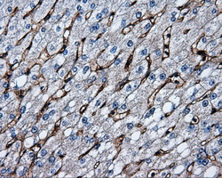
Immunohistochemical staining of paraffin-embedded liver tissue within the normal limits using anti-CD36mouse monoclonal antibody. (Heat-induced epitope retrieval by 10mM citric buffer, pH6.0, 100°C for 10min, Dilution 1:50)IHC
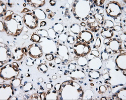
Immunohistochemical staining of paraffin-embedded Kidney tissue within the normal limits using anti-CD36mouse monoclonal antibody. (Heat-induced epitope retrieval by 10mM citric buffer, pH6.0, 100°C for 10min, Dilution 1:50)IHC
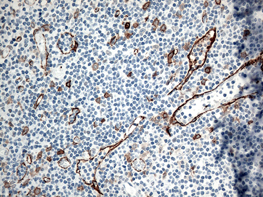
Immunohistochemical staining of paraffin-embedded Human tonsil within the normal limits using anti-CD36 mouse monoclonal antibody. (Heat-induced epitope retrieval by 1mM EDTA in 10mM Tris buffer (pH8.5) at 120°C for 3min) (1:500)IHC
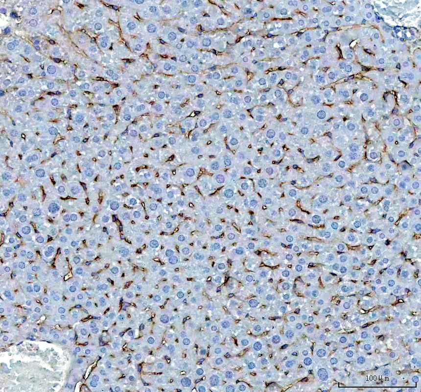
IHC analysis of CD36 using anti-CD36 antibody. CD36 was detected in a paraffin-embedded section of mouse liver tissue. The tissue section was incubated with mouse anti-CD36 Antibody at a dilution of 1:200 and developed using HRP Conjugated mouse IgG Super Vision Assay Kit with DAB as the chromogen.ICC/IF
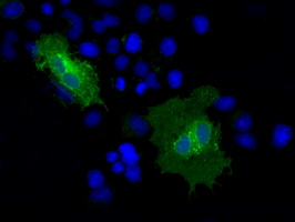
Anti-CD36 mouse monoclonal antibody immunofluorescent staining of COS7 cells transiently transfected by pCMV6-ENTRY CD36.FC
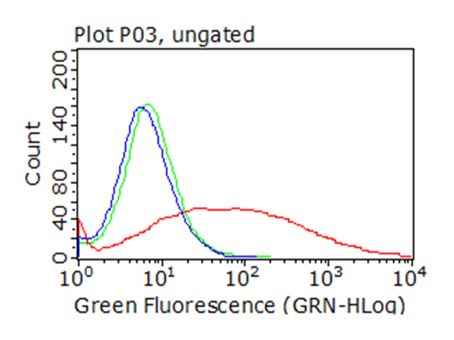
HEK293T cells transfected with either overexpress plasmid (Red), compared to an IgG isotype control, (Green) or empty vector control plasmid (Blue) were immunostained by anti-CD36 antibody, and then analyzed by flow cytometry (1:100).| Product Name | Anti-CD36 Antibody |
|---|---|
| Antibody Type | Primary Antibodies |
| Immunogen | Full length human recombinant protein of human CD36 (NP_000063) produced in HEK293T cell. |
| Clonality | Monoclonal |
|---|---|
| Isotype | IgG1 |
| Host Species | Mouse |
| Tested Applications | FCICC/IFIHCWB |
| WB:1:2000 IHC:1:50 ICC/IF:1:100 FC:1:100 |
|
| Species Reactivity | HumanMouseRat |
| Concentration | 1mg/ml |
| Purification | Protein A/G |
| Gene Symbol | CD36 |
|---|---|
| Gene Synonyms | FAT GP4 GP3B GPIV CHDS7 PASIV SCARB3 BDPLT10 |
| Gene Full Name | CD36 molecule (CD36 blood group) |
| Gene Summary | The protein encoded by this gene is the fourth major glycoprotein of the platelet surface and serves as a receptor for thrombospondin in platelets and various cell lines. Since thrombospondins are widely distributed proteins involved in a variety of adhesive processes, this protein may have important functions as a cell adhesion molecule. It binds to collagen, thrombospondin, anionic phospholipids and oxidized LDL. It directly mediates cytoadherence of Plasmodium falciparum parasitized erythrocytes and it binds long chain fatty acids and may function in the transport and/or as a regulator of fatty acid transport. Mutations in this gene cause platelet glycoprotein deficiency. Multiple alternatively spliced transcript variants have been found for this gene. [provided by RefSeq, Feb 2014] |
| Alternative Names | CD36 CHDS7 FAT Fatty acid translocase Glycoprotein IIIb GP3B GP4 GPIIIB GPIV PAS 4 PAS IV PASIV Platelet glycoprotein 4 Platelet glycoprotein IV SCARB3 Thrombospondin receptor |
| Molecular Weight(MW) | 53kDa(Observed MW 88kDa) |
| Cellular Localization | Cell membrane |
WB
Western blot analysis of anti-CD36 antibody. The sample well of each lane was loaded with 30 ug of sample under reducing conditions. Lane 1: human placenta tissue lysates. After electrophoresis, proteins were transferred to a membrane. Then the membrane was incubated with mouse anti-CD36 antigen affinity purified monoclonal antibody and probed with a goat anti-mouse IgG-HRP secondary antibody. The signal is developed using ECL Plus Western Blotting Substrate.
IHC
Immunohistochemical staining of paraffin-embedded liver tissue within the normal limits using anti-CD36mouse monoclonal antibody. (Heat-induced epitope retrieval by 10mM citric buffer, pH6.0, 100°C for 10min, Dilution 1:50)
IHC
Immunohistochemical staining of paraffin-embedded Kidney tissue within the normal limits using anti-CD36mouse monoclonal antibody. (Heat-induced epitope retrieval by 10mM citric buffer, pH6.0, 100°C for 10min, Dilution 1:50)
IHC
Immunohistochemical staining of paraffin-embedded Human tonsil within the normal limits using anti-CD36 mouse monoclonal antibody. (Heat-induced epitope retrieval by 1mM EDTA in 10mM Tris buffer (pH8.5) at 120°C for 3min) (1:500)
IHC
IHC analysis of CD36 using anti-CD36 antibody. CD36 was detected in a paraffin-embedded section of mouse liver tissue. The tissue section was incubated with mouse anti-CD36 Antibody at a dilution of 1:200 and developed using HRP Conjugated mouse IgG Super Vision Assay Kit with DAB as the chromogen.
ICC/IF
Anti-CD36 mouse monoclonal antibody immunofluorescent staining of COS7 cells transiently transfected by pCMV6-ENTRY CD36.
FC
HEK293T cells transfected with either overexpress plasmid (Red), compared to an IgG isotype control, (Green) or empty vector control plasmid (Blue) were immunostained by anti-CD36 antibody, and then analyzed by flow cytometry (1:100).| Application Notes | WB:1:2000 IHC:1:50 ICC/IF:1:100 FC:1:100 |
|---|
| Form | Liquid |
|---|---|
| Storage Instructions | Stable for 12 months from date of receipt. Store at -20°C as received. |
| Storage Buffer | PBS (pH 7.3) containing 1% BSA, 50% glycerol and 0.02% sodium azide. |


