WB
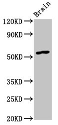
Western Blot, Positive WB detected in: Rat brain tissue, All lanes: GDF10 antibody at 4.5μg/ml, Secondary, Goat polyclonal to rabbit IgG at 1/50000 dilution.IHC
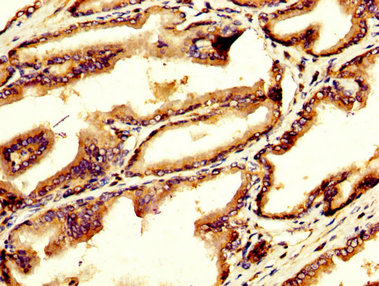
IHC image of GDF10 Antibody diluted at 1:200 and staining in paraffin-embedded human prostate tissue performed on a Leica BondTM system. After dewaxing and hydration, antigen retrieval was mediated by high pressure in a citrate buffer (pH 6.0). Section was blocked with 10% normal goat serum 30min at RT. Then primary antibody (1% BSA) was incubated at 4°C overnight. The primary is detected by a biotinylated secondary antibody and visualized using an HRP conjugated SP system.| Product Name | GDF10 Antibody |
|---|---|
| Antibody Type | Primary Antibodies |
| Immunogen | Recombinant Human Growth/differentiation factor 10 protein (369-478AA) |
| Clonality | Polyclonal |
|---|---|
| Isotype | IgG |
| Host Species | Rabbit |
| Tested Applications | IHCWB |
| WB:1:500-1:2000 IHC:1:20-1:200 |
|
| Species Reactivity | HumanRat |
| Concentration | 1mg/ml |
| Purification | Protein G |
| Gene Symbol | GDF10 |
|---|---|
| Gene Synonyms | BIP BMP3B BMP-3b |
| Gene Full Name | growth differentiation factor 10 |
| Gene Summary | This gene encodes a secreted ligand of the TGF-beta (transforming growth factor-beta) superfamily of proteins. Ligands of this family bind various TGF-beta receptors leading to recruitment and activation of SMAD family transcription factors that regulate gene expression. The encoded preproprotein is proteolytically processed to generate each subunit of the disulfide-linked homodimer. This promotes neural repair after stroke. Additionally, this protein may act as a tumor suppressor and reduced expression of this gene is associated with oral cancer. [provided by RefSeq, Jul 2016] |
| Molecular Weight(MW) | 54kDa(Observed MW 54kDa) |
| Cellular Localization | Secreted. |
WB
Western Blot, Positive WB detected in: Rat brain tissue, All lanes: GDF10 antibody at 4.5μg/ml, Secondary, Goat polyclonal to rabbit IgG at 1/50000 dilution.
IHC
IHC image of GDF10 Antibody diluted at 1:200 and staining in paraffin-embedded human prostate tissue performed on a Leica BondTM system. After dewaxing and hydration, antigen retrieval was mediated by high pressure in a citrate buffer (pH 6.0). Section was blocked with 10% normal goat serum 30min at RT. Then primary antibody (1% BSA) was incubated at 4°C overnight. The primary is detected by a biotinylated secondary antibody and visualized using an HRP conjugated SP system.| Application Notes | WB:1:500-1:2000 IHC:1:20-1:200 |
|---|
| Form | Liquid |
|---|---|
| Storage Instructions | Upon receipt, store at -20°C or -80°C. Avoid repeated freeze. |
| Storage Buffer | Preservative: 0.03% Proclin 300 Constituents: 50% Glycerol, 0.01M PBS, pH 7.4 |


