DB
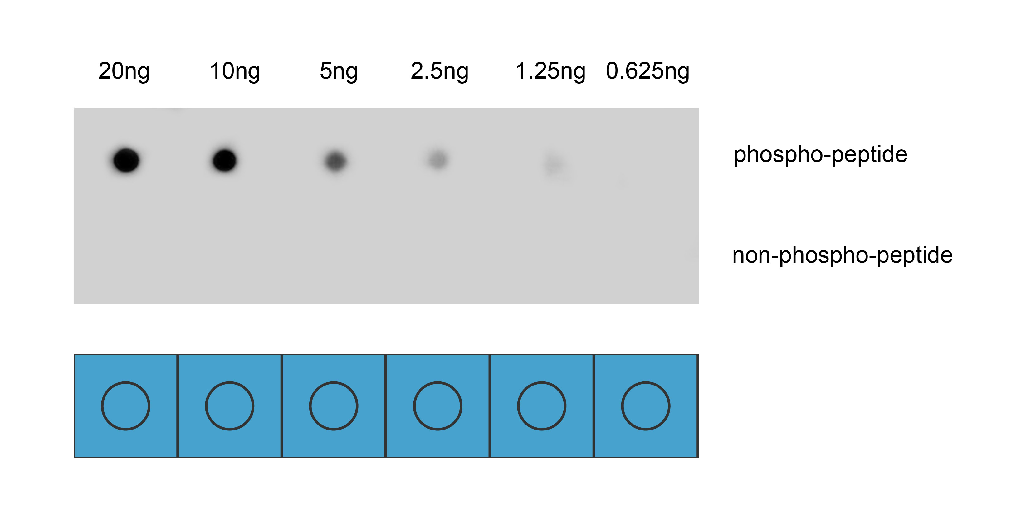
Dot blot analysis was performed using Anti-Phospho-NuMA(Ser395) antibody to detect two peptide samples: lane 1 contains the phospho-peptide , and lane 2 contains the non-phospho-peptide. Transfer the proteins onto a NC membrane (OM790001), and block it with TBST (OM750016) containing 5% BSA(OM297470) for 1 hour. Dilute the primary antibody with the antibody diluent (OM750012) at a ratio of 1:1000, and incubate it at room temperature for 2 hours. Wash the membrane 3 times with TBST (OM750016), 5 minutes each time. At room temperature, dilute the secondary antibody, Goat Anti-Rabbit IgG(H&L)-HRP (OM643487), at a ratio of 1:10000 and incubate for 1 hour. Wash the membrane 3 times with TBST (OM750016) again, 5 minutes each time. Use ECL (OM625701) for luminescence. Exposure time: 30 s.ELISA
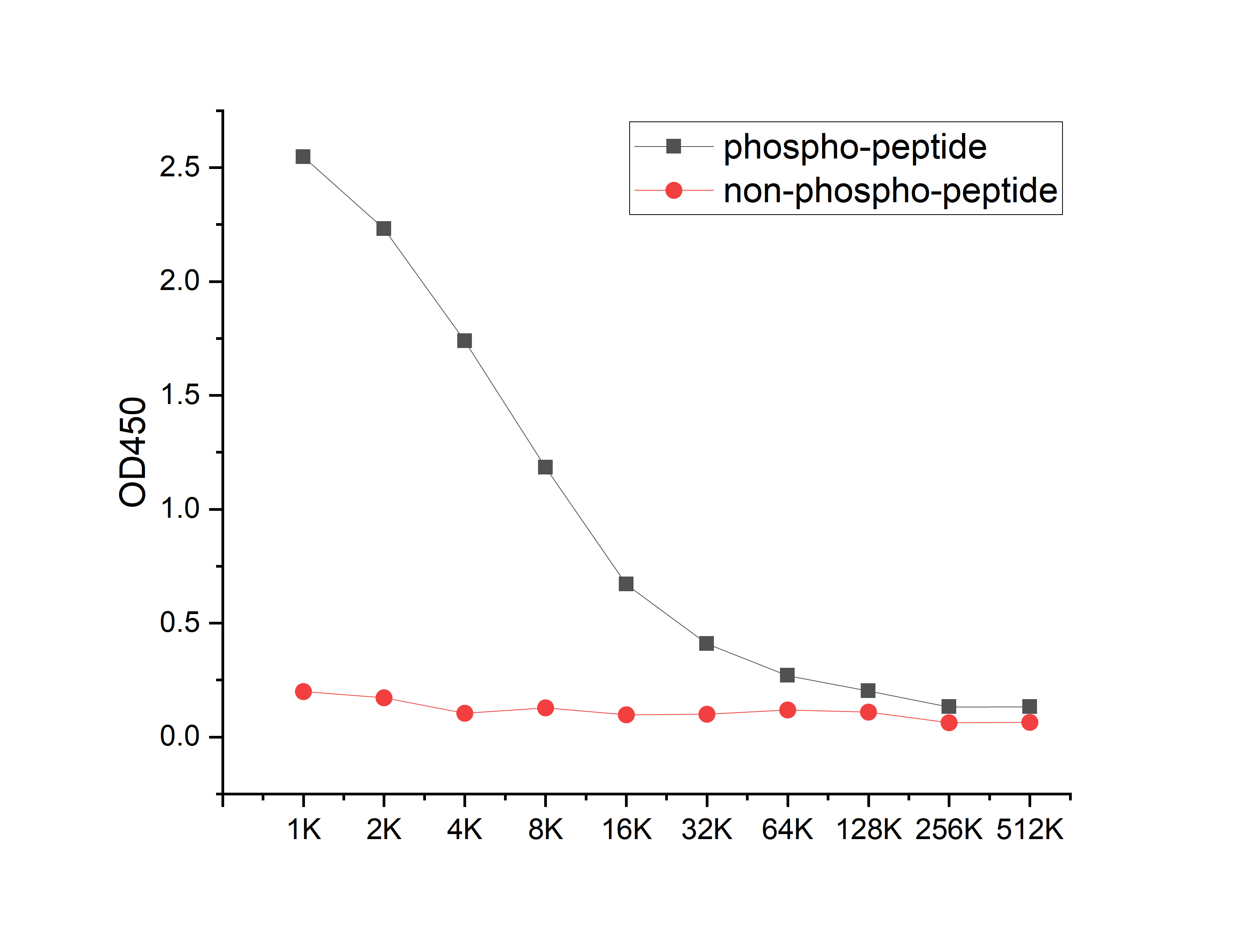
Coat the plate with 100ng immunizing antigen. The phospho-specific antibody,pbs only can specifically recognize phosphorylated peptide and enable quantitative detection over a wide dynamic range. Non-phosphorylated peptide shows no cross-reactivity, and the background signal remains low, indicating that the phospho-specific antibody,pbs only possesses high specificity and an excellent signal-to-noise ratio. Black line: phospho-peptide; Rad line: non-phospho peptide| Product Name | Anti-Phospho-NuMA(Ser395) antibody |
|---|---|
| Antibody Type | Primary Antibodies |
| Immunogen | Synthetic peptide conjugated with KLH |
| Modification | p-Ser395 |
| Clonality | Polyclonal |
|---|---|
| Isotype | IgG |
| Host Species | Rabbit |
| Tested Applications | DBELISA |
| DB:1:1000 |
|
| Concentration | 1mg/ml |
| Purification | Affinity purified |
| Gene Symbol | NUMA1 |
|---|---|
| Gene Synonyms | NUMA, NMP-22 |
| Gene Full Name | nuclear mitotic apparatus protein 1 |
| Gene Summary | This gene encodes a large protein that forms a structural component of the nuclear matrix. The encoded protein interacts with microtubules and plays a role in the formation and organization of the mitotic spindle during cell division. Chromosomal translocation of this gene with the RARA (retinoic acid receptor, alpha) gene on chromosome 17 have been detected in patients with acute promyelocytic leukemia. Alternative splicing results in multiple transcript variants. [provided by RefSeq, Nov 2013] |
| Alternative Names | Centrophilin stabilizes mitotic spindle in mitotic cells NMP 22 Nuclear matrix protein 22 Nuclear mitotic apparatus protein 1 Nuclear mitotic apparatus protein NUMA 1 NUMA NuMA protein NUMA1 NUMA1_HUMAN SP H antigen SP-H antigen Structural nuclear protein |
| Molecular Weight(MW) | 238 kDa |
| Cellular Localization | Nucleus. Nucleus>Nucleoplasm. Nucleus matrix. Chromosome. Cytoplasm>Cytoskeleton. Cytoplasm>Cytoskeleton>Microtubule organizing center>Centrosome. Cytoplasm>Cytoskeleton>Spindle pole. Cytoplasm>Cell cortex. Cell membrane>Lipid-anchor>Cytoplasmic side. Lateral cell membrane. |
| SwissProt ID | Q14980 |
|---|
DB
Dot blot analysis was performed using Anti-Phospho-NuMA(Ser395) antibody to detect two peptide samples: lane 1 contains the phospho-peptide , and lane 2 contains the non-phospho-peptide. Transfer the proteins onto a NC membrane (OM790001), and block it with TBST (OM750016) containing 5% BSA(OM297470) for 1 hour. Dilute the primary antibody with the antibody diluent (OM750012) at a ratio of 1:1000, and incubate it at room temperature for 2 hours. Wash the membrane 3 times with TBST (OM750016), 5 minutes each time. At room temperature, dilute the secondary antibody, Goat Anti-Rabbit IgG(H&L)-HRP (OM643487), at a ratio of 1:10000 and incubate for 1 hour. Wash the membrane 3 times with TBST (OM750016) again, 5 minutes each time. Use ECL (OM625701) for luminescence. Exposure time: 30 s.
ELISA
Coat the plate with 100ng immunizing antigen. The phospho-specific antibody,pbs only can specifically recognize phosphorylated peptide and enable quantitative detection over a wide dynamic range. Non-phosphorylated peptide shows no cross-reactivity, and the background signal remains low, indicating that the phospho-specific antibody,pbs only possesses high specificity and an excellent signal-to-noise ratio. Black line: phospho-peptide; Rad line: non-phospho peptide| Application Notes | DB:1:1000 |
|---|
| Form | Liquid |
|---|---|
| Storage Instructions | Shipped at 4°C. Store at +4°C short term (1-2 weeks). Store at -20°C long term. Avoid freeze / thaw cycle. |
| Storage Buffer | Purified antibody in PBS with 0.05% sodium azide,40% Glycerol. |


