ELISA
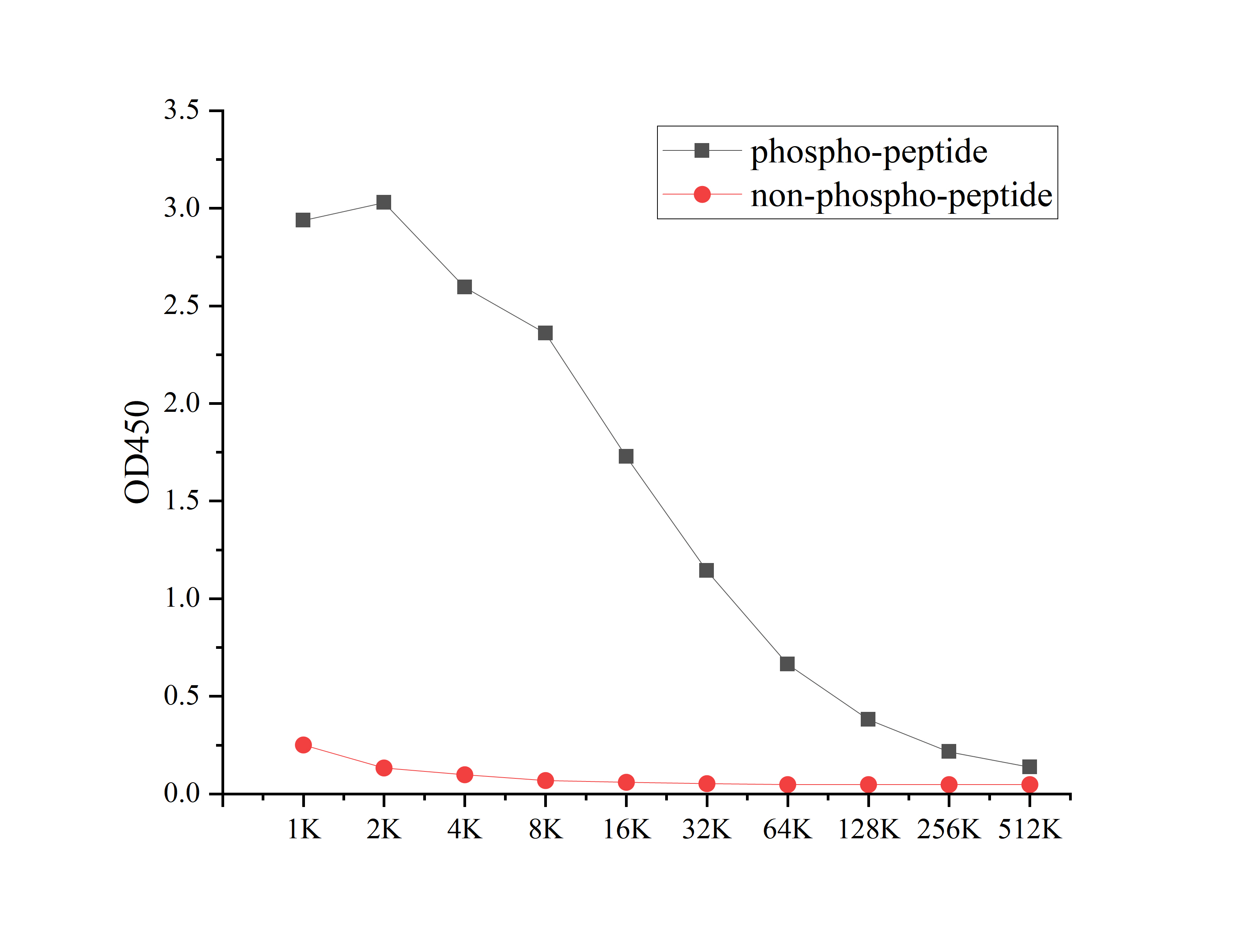
Coat the plate with 100ng immunizing antigen. The phospho-specific antibody,pbs only can specifically recognize phosphorylated peptide and enable quantitative detection over a wide dynamic range. Non-phosphorylated peptide shows no cross-reactivity, and the background signal remains low, indicating that the phospho-specific antibody,pbs only possesses high specificity and an excellent signal-to-noise ratio. Black line: phospho-peptide; Rad line: non-phospho peptideDB
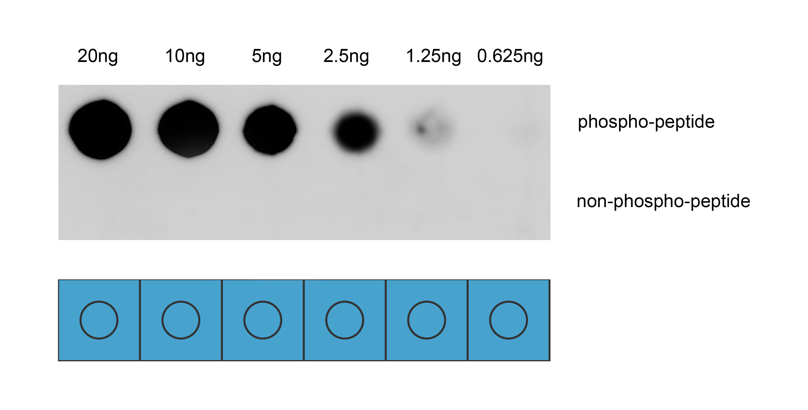
Dot blot analysis was performed using Anti-Phospho-VE-Cadherin(Tyr731) antibody to detect two peptide samples: lane 1 contains the phospho-peptide , and lane 2 contains the non-phospho-peptide. Transfer the proteins onto a NC membrane (OM790001), and block it with TBST (OM750016) containing 5% BSA(OM297470) for 1 hour. Dilute the primary antibody with the antibody diluent (OM750012) at a ratio of 1:1000, and incubate it at room temperature for 2 hours. Wash the membrane 3 times with TBST (OM750016), 5 minutes each time. At room temperature, dilute the secondary antibody, Goat Anti-Rabbit IgG(H&L)-HRP (OM643487), at a ratio of 1:10000 and incubate for 1 hour. Wash the membrane 3 times with TBST (OM750016) again, 5 minutes each time. Use ECL (OM625701) for luminescence. Exposure time: 30 s.| Product Name | Anti-Phospho-VE-Cadherin (Tyr731) antibody |
|---|---|
| Antibody Type | Primary Antibodies |
| Immunogen | Synthetic peptide conjugated with KLH. |
| Modification | p-Tyr731 |
| Clonality | Polyclonal |
|---|---|
| Isotype | IgG |
| Host Species | Rabbit |
| Tested Applications | DBELISA |
| DB:1:1000 |
|
| Concentration | 1mg/ml |
| Purification | Affinity purified |
| Gene Symbol | CDH5 |
|---|---|
| Gene Synonyms | 7B4 CD144 |
| Gene Full Name | cadherin 5 |
| Gene Summary | This gene encodes a classical cadherin of the cadherin superfamily. The encoded preproprotein is proteolytically processed to generate the mature glycoprotein. This calcium-dependent cell-cell adhesion molecule is comprised of five extracellular cadherin repeats, a transmembrane region and a highly conserved cytoplasmic tail. Functioning as a classical cadherin by imparting to cells the ability to adhere in a homophilic manner, this protein plays a role in endothelial adherens junction assembly and maintenance. This gene is located in a gene cluster in a region on the long arm of chromosome 16 that is involved in loss of heterozygosity events in breast and prostate cancer. [provided by RefSeq, Nov 2015] |
| Alternative Names | 7B 4 7B4 7B4 antigen CADH5_HUMAN Cadherin 5 Cadherin 5 type 2 Cadherin 5 type 2 (vascular endothelium) Cadherin 5 type 2 VE cadherin (vascular epithelium) cadherin vascular endothelial cadherin vascular endothelial 1 Cadherin-5 Cadherin5 CD 144 CD144 CD144 antigen CDH 5 CDH5 CDH5 protein Endothelial specific cadherin FLJ17376 OTTHUMP00000174777 Vascular endothelial cadherin Vascular epithelium cadherin VE Cad VE-cadherin VEC |
| Molecular Weight(MW) | 88 kDa |
| Cellular Localization | Cell junction, Cell membrane, Cytoplasm, Membrane. |
| SwissProt ID | P33151 |
|---|
ELISA
Coat the plate with 100ng immunizing antigen. The phospho-specific antibody,pbs only can specifically recognize phosphorylated peptide and enable quantitative detection over a wide dynamic range. Non-phosphorylated peptide shows no cross-reactivity, and the background signal remains low, indicating that the phospho-specific antibody,pbs only possesses high specificity and an excellent signal-to-noise ratio. Black line: phospho-peptide; Rad line: non-phospho peptide
DB
Dot blot analysis was performed using Anti-Phospho-VE-Cadherin(Tyr731) antibody to detect two peptide samples: lane 1 contains the phospho-peptide , and lane 2 contains the non-phospho-peptide. Transfer the proteins onto a NC membrane (OM790001), and block it with TBST (OM750016) containing 5% BSA(OM297470) for 1 hour. Dilute the primary antibody with the antibody diluent (OM750012) at a ratio of 1:1000, and incubate it at room temperature for 2 hours. Wash the membrane 3 times with TBST (OM750016), 5 minutes each time. At room temperature, dilute the secondary antibody, Goat Anti-Rabbit IgG(H&L)-HRP (OM643487), at a ratio of 1:10000 and incubate for 1 hour. Wash the membrane 3 times with TBST (OM750016) again, 5 minutes each time. Use ECL (OM625701) for luminescence. Exposure time: 30 s.| Application Notes | DB:1:1000 |
|---|
| Form | Liquid |
|---|---|
| Storage Instructions | Store at 4°C short term. Aliquot and store at -20°C long term. Avoid freeze/thaw cycles. |
| Storage Buffer | Purified antibody in PBS with 0.05% sodium azide,40% Glycerol. |


