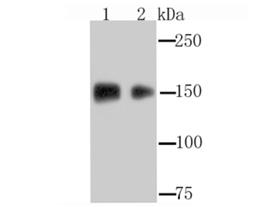
Fig1: Western blot analysis of EGFR on A431 (1) and HepG2 (2) cell lysate using anti-EGFR antibody at 1/1,000 dilution.
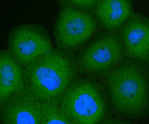
Fig2: ICC staining EGFR in A431 cells (green). The nuclear counter stain is DAPI (blue). Cells were fixed in paraformaldehyde, permeabilised with 0.25% Triton X100/PBS.
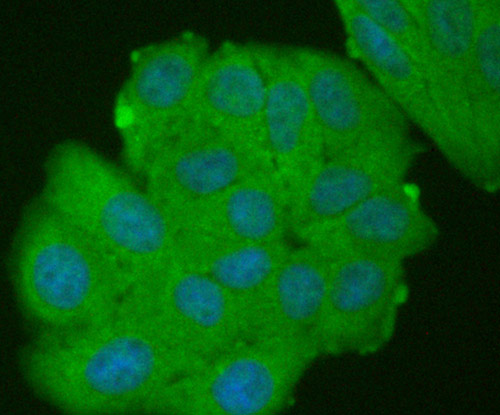
Fig3: ICC staining EGFR in HepG2 cells (green). The nuclear counter stain is DAPI (blue). Cells were fixed in paraformaldehyde, permeabilised with 0.25% Triton X100/PBS.
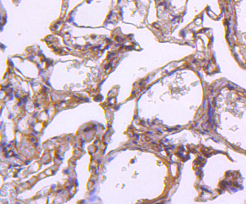
Fig4: Immunohistochemical analysis of paraffin-embedded human lung tissue using anti-EGFR antibody. Counter stained with hematoxylin.
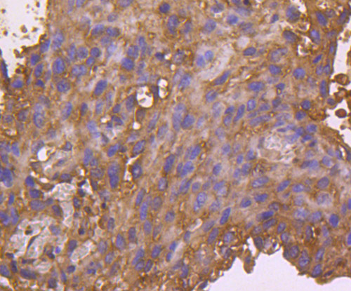
Fig5: Immunohistochemical analysis of paraffin-embedded human breast cancer tissue using anti-EGFR antibody. Counter stained with hematoxylin.
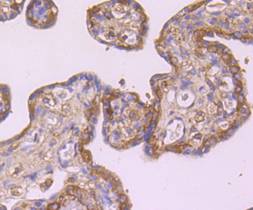
Fig6: Immunohistochemical analysis of paraffin-embedded human placenta tissue using anti-EGFR antibody. Counter stained with hematoxylin.
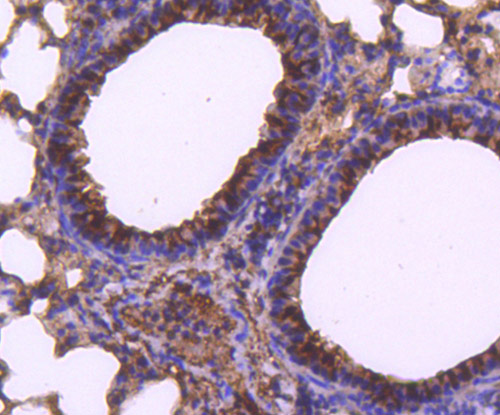
Fig7: Immunohistochemical analysis of paraffin-embedded mouse lung tissue using anti-EGFR antibody. Counter stained with hematoxylin.
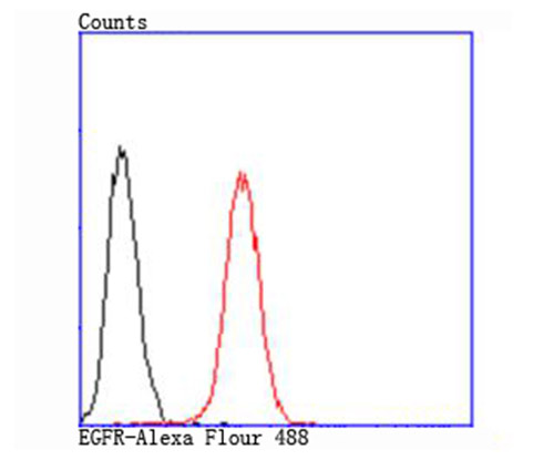
Fig8: Flow cytometric analysis of PANC-1 cells with EGFR antibody at 1/100 dilution (red) compared with an unlabelled control (cells without incubation with primary antibody; black).
Product Profile
| Product Name | EGFR |
|---|---|
| Antibody Type | Primary Antibodies |
| Product description |
|
| Immunogen | Recombinant protein. |
Key Feature
| Clonality | Polyclonal |
|---|---|
| Isotype | IgG |
| Host Species | Rabbit |
| Tested Applications | |
WB:1:500~1:2000 ICC:1:200~1:500 IHC:1:50~1:200 FC:1:200~1:500 Notes:Optimal dilutions/concentrations should be determined by the researcher. |
|
| Species Reactivity | |
| Concentration | 1 mg/mL. |
Target Information
| Alternative Names | Avian erythroblastic leukemia viral (v erb b) oncogene homolog antibody
Cell growth inhibiting protein 40 antibody
Cell proliferation inducing protein 61 antibody
EGF R antibody
EGFR antibody
EGFR_HUMAN antibody
Epidermal growth factor receptor (avian erythroblastic leukemia viral (v erb b) oncogene homolog) antibody
Epidermal growth factor receptor (erythroblastic leukemia viral (v erb b) oncogene homolog avian) antibody
Epidermal growth factor receptor antibody
erb-b2 receptor tyrosine kinase 1 antibody
ERBB antibody
ERBB1 antibody
Errp antibody
HER1 antibody
mENA antibody
NISBD2 antibody
Oncogen ERBB antibody
PIG61 antibody
Proto-oncogene c-ErbB-1 antibody
Receptor tyrosine protein kinase ErbB 1 antibody
Receptor tyrosine-protein kinase ErbB-1 antibody
SA7 antibody
Species antigen 7 antibody
Urogastrone antibody
v-erb-b Avian erythroblastic leukemia viral oncogen homolog antibody
wa2 antibody
Wa5 antibody
|
|---|---|
| Molecular Weight(MW) | 175 kDa |
| Cellular Localization | Secreted and Cell membrane. Endosome membrane. Nucleus. |
Database Links
| SwissProt ID | P00533 Q01279 |
|---|
Application
-

Application
Fig1: Western blot analysis of EGFR on A431 (1) and HepG2 (2) cell lysate using anti-EGFR antibody at 1/1,000 dilution.
-

Application
Fig2: ICC staining EGFR in A431 cells (green). The nuclear counter stain is DAPI (blue). Cells were fixed in paraformaldehyde, permeabilised with 0.25% Triton X100/PBS.
-

Application
Fig3: ICC staining EGFR in HepG2 cells (green). The nuclear counter stain is DAPI (blue). Cells were fixed in paraformaldehyde, permeabilised with 0.25% Triton X100/PBS.
-

Application
Fig4: Immunohistochemical analysis of paraffin-embedded human lung tissue using anti-EGFR antibody. Counter stained with hematoxylin.
-

Application
Fig5: Immunohistochemical analysis of paraffin-embedded human breast cancer tissue using anti-EGFR antibody. Counter stained with hematoxylin.
-

Application
Fig6: Immunohistochemical analysis of paraffin-embedded human placenta tissue using anti-EGFR antibody. Counter stained with hematoxylin.
-

Application
Fig7: Immunohistochemical analysis of paraffin-embedded mouse lung tissue using anti-EGFR antibody. Counter stained with hematoxylin.
-

Application
Fig8: Flow cytometric analysis of PANC-1 cells with EGFR antibody at 1/100 dilution (red) compared with an unlabelled control (cells without incubation with primary antibody; black).
| Positive Control | A431, HepG2, PANC-1, human lung tissue, human breast cancer tissue, human placenta tissue, mouse lung tissue. |
|---|---|
| Application Notes | WB:1:500~1:2000 ICC:1:200~1:500 IHC:1:50~1:200 FC:1:200~1:500 Notes:Optimal dilutions/concentrations should be determined by the researcher. |
Additional Information
| Form | Liquid |
|---|---|
| Storage Instructions | Store at +4℃ after thawing. Aliquot store at -20℃ or -80℃. Avoid repeated freeze / thaw cycles. |
| Storage Buffer | 1*TBS (pH7.4), 0.5%BSA, 50%Glycerol. Preservative: 0.05% Sodium Azide. |
- Related products
- Camel Growth Hormone (GH) ELISA Kit OM642709
- Camel Insulin-like growth factors 1 (IGF-1) ELISA Kit OM642708
- Human HIV-1 p24 core protein (HIV-1 p24) antibody ELISA Kit OM642706
- Antigen Repair 20X pH6.0 OM642703
- 647 labeled Tyramide OM642699
-
- ASSAY KITS
-
- SERUM
2013 © Omnimabs , All Rights Reserved.

